Date
TSH (mIU/L)
Tg (ng/ml)
ANTI-Tg-AB (IU/ml)
Course
10/04/13
4.18
16,340
<2
Thyroidectomy 9/7/13
10/26/13
1.09
12,503
<2
IMRT to lumbar spine 11/13
12/22/13
0.36
9517
<2
120 mCi 131-I (12/23/13)
01/13/14
2.99
3303
<2
02/23/14
3.72
1662
<20 (different assay)
03/12/14
54
2840
<2
360 mCi 131-I (3/17/14)
06/02/14
1.92
142
<2
08/22/14
0.09
64
<2
Assessment
This 55-year-old man presented with widespread metastases to multiple bones which were subsequently confirmed to be from a follicular carcinoma of the thyroid. According to his medical record notes, his serum thyroglobulin levels were quite high and fell after surgery and radioiodine ablation (Values not available Table 28.1), but remained markedly elevated. His post-therapy scan indicates fairly good radioiodine uptake suggesting potential benefit from additional radioactive iodine treatment. Our approach will be to do additional imaging to assess the current extent and location of residual disease in order to determine possible therapeutic approaches.
Relevant Literature
In general, distant metastases from thyroid cancer can be seen in 4–6 % of patients at initial presentation [1, 2]. Distant metastatic disease can subsequently develop in 10–30 % of patients [3, 4], depending on the histology (about 10 % in papillary cancer and 20–30 % in follicular and Hurthle cell cancers), most commonly to the lung and bones. The presence of distant metastases may be heralded by an extremely elevated serum thyroglobulin, with the findings confirmed by plain radiography, 131-I whole body scan, CT, MRI, PET/CT, or bone scan. Radioiodine uptake in a bone lesion will indicate that it is derived from thyroid cancer, but absent that, a bone biopsy is advisable to confirm the diagnosis. Other imaging modalities for bone lesions may include thallium-201, technetium-99m, or iodine-124 PET. Survival with metastatic disease will depend upon the site of the metastases, the age of the patient, and whether or not the lesions are radioiodine avid. Other factors influencing prognosis and statistically significant by univariate analysis include gender, extent of surgery, and histologic type [1, 3]. Clinically, patients will present with bone pain or fracture, swelling at the site, or spinal metastases with symptoms of cord compression. The more common bony sites for metastases are the spine, pelvis, ribs, femur, and skull. Bone metastases connote a significantly poorer prognosis than that noted in the majority of thyroid cancer patients. In a retrospective analysis by Shoup et al. [5] of 242 patients, 40 % had metastases on presentation and they had a 10-year survival of 26 % with a median survival of 4.1 years. The importance of age is apparent as seen with a 10-year survival in those patients <45 years old of 58 % compared to only 13 % in those >45 years old.
The treatment of bone metastases typically involves radioiodine, assuming that the lesions are RAI avid, and it is the younger patients who are more likely to have RAI uptake, a fact that likely is linked to their better survival statistics. This may account for why some studies show clear benefit of RAI therapy [6, 7], while others do not [8, 9]. Preparation for radioiodine therapy requires an elevated serum TSH level which may be achieved by either thyroid hormone withdrawal or administration of recombinant human TSH [10, 11]. Those lesions that are FDG/PET avid tend to be less differentiated and more resistant to RAI treatment [12, 13]. RAI may be administered as either a standard or “empiric” dose or activity based upon consensus guidelines and physician experience, or as a “dosimetric” activity based on how the patient’s body handles (retains and excretes) a given activity of radioiodine. When there is RAI uptake, arguments have been made for administering the highest dose feasible that is within safe limits as can be determined by dosimetry [14], and dosimetric approaches have been shown to be potentially more efficacious than empiric dosage [15]. However, relatively lower doses appear to be appropriate in older patients [16]. In the absence of RAI uptake, other therapeutic modalities may be tried but will be associated with less salutary effect. There is hope that redifferentiating agents such as selumetinib might prove useful for these patients in the future [17]. When feasible, particularly for isolated and/or symptomatic bone metastasis, other approaches to management could include surgical resection with or without cementoplasty, radiofrequency ablation, external radiation, arterial embolization, or targeted chemotherapy, along with adjunctive use of bisphosphonates [18]. Pak et al. [19] reviewed a 32-year experience with surgical metastasectomy with 51 metastases excised in 29 patients who had a variety of types of thyroid cancer. Many of the patients received adjunctive therapy with XRT and RAI as well with a resultant 78 % survival at 5 years that dropped to 50 % at 10 years, with age >45 a poor prognostic factor.
External beam radiation is employed for pain relief and for lesions in critical or weight-bearing areas, as well as for those lesions not amenable to surgery or RAI treatment. It is typically given in doses of 50 Gy in 25 fractions for solitary bone lesions and 40 Gy in 20 fractions for vertebral lesions. Arterial embolization has been employed by Eustatia-Rutten and colleagues [20] with 41 embolizations in 16 patients resulting in clinical improvement in 60 % of subjects. The procedure requires visualization of the artery feeding the metastasis by selective catheterization, and then particles of isobutyl, Gelfoam, or polyvinyl alcohol are injected with relief of pain and improvement in neurologic symptoms. The best results are seen in those patients who also have had RAI, XRT, or surgical treatment. Radiofrequency ablation (RFA) [21, 22] uses probes inserted under CT guidance to generate thermal energy to achieve local tissue temperatures >50 °C inducing cell death. RFA has rarely been applied to bone lesions and has found greater application to lesions in the liver, kidney, or bone. While planar radioiodine scanning with iodine-131 is the primary imaging procedure for differentiated thyroid cancer, scanning with iodine-124 PET offers advantages of detecting both positron emission as well as the spatial distribution of radioiodine uptake and allows improved detection of the extent of residual disease [23–27].
Subsequent Management
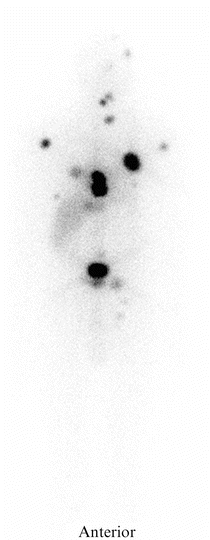
The degree of thyroglobulin elevation indicated persistent residual disease. Given that his earlier post-therapy radioiodine scan showed significant uptake and the actual activity administered (120 mCi) was relatively low, the patient was deemed a good candidate for higher-dose radioiodine therapy to be determined by dosimetry [14]. He was withdrawn from his levothyroxine therapy, and after 3 weeks, his serum TSH level had risen to 42 mU/L. He then underwent routine total body scanning with iodine-131 (Fig. 28.1) as well as iodine-124 PET scanning which again clearly delineated good isotope uptake, with particularly better visualization of the extent of bone metastases by the iodine-124 PET images (Fig. 28.2). Regional lesional dosimetry afforded by iodine-124 indicated that sufficient uptake of activity in specific lesions could be achieved for therapeutic benefit based on earlier data on locoregional dosimetry [25, 28]. The dosimetry calculations predicted that he could receive as much as 410 mCi of iodine-131 safely and have less than 100 mCi retained in total body at 48 h. He was treated with 360 mCi iodine-131 in late March 2014 at a time that his TSH measured 54 mU/L; the post-therapy scan was essentially identical to the pretreatment diagnostic scan demonstrating good uptake in multiple sites estimated to approximate 72 Gy or 7200 rads. Follow-up studies by his physicians in West Virginia on June 2, 2014, included a serum thyroglobulin of 142 ng/ml and a TSH of 1.92 mU/L. His dose of levothyroxine was increased, and the last studies recorded to date on August 22, 2014 included a thyroglobulin of 64 ng/ml and a TSH of 0.09 mU/L. His total white blood cell, absolute neutrophil, and platelet counts have remained within normal limits. Recommendations to his physicians included continuing suppressive dosage of levothyroxine, adjunctive therapy with bisphosphonates or denosumab, monitoring serum thyroglobulin every 6 months, considering local intervention on selective lesions by XRT or RFA as warranted, and a potential additional dosimetric radioiodine dose based upon demonstrated progressive disease, persistent iodine uptake, and relatively normal blood counts.
 A Case of Multifocal Papillary Thyroid Microcarcinoma
A Patient with Papillary Thyroid Cancer and Biochemical Evidence of Disease at the One-Year Follow-Up Visit
A Case of Papillary Thyroid Cancer Without Aggressive Histological Features with Nodal Metastases in an Older Patient
RAI-Refractory, Advanced Differentiated Thyroid Cancer Receiving Tyrosine Kinase Inhibitor Treatment: Checking for Drug-Drug Interactions
A Case of Multifocal Papillary Thyroid Microcarcinoma
A Patient with Papillary Thyroid Cancer and Biochemical Evidence of Disease at the One-Year Follow-Up Visit
A Case of Papillary Thyroid Cancer Without Aggressive Histological Features with Nodal Metastases in an Older Patient
RAI-Refractory, Advanced Differentiated Thyroid Cancer Receiving Tyrosine Kinase Inhibitor Treatment: Checking for Drug-Drug Interactions
 A Child with Papillary Thyroid Cancer and Locally Advanced Disease but No Distant Metastasis
 Timing and Extent of Surgery for a Pediatric Patient with Hereditary MTC and Positive Screening for the S891A RET Mutation


Related posts:
A Case of Multifocal Papillary Thyroid Microcarcinoma
A Patient with Papillary Thyroid Cancer and Biochemical Evidence of Disease at the One-Year Follow-Up Visit
A Case of Papillary Thyroid Cancer Without Aggressive Histological Features with Nodal Metastases in an Older Patient
RAI-Refractory, Advanced Differentiated Thyroid Cancer Receiving Tyrosine Kinase Inhibitor Treatment: Checking for Drug-Drug Interactions
A Child with Papillary Thyroid Cancer and Locally Advanced Disease but No Distant Metastasis
Timing and Extent of Surgery for a Pediatric Patient with Hereditary MTC and Positive Screening for the S891A RET Mutation
Stay updated, free articles. Join our Telegram channel
Full access? Get Clinical Tree
