Fig. 5.1
Rundle’s curve depicting the clinical course of TED demonstrating disease activity (dotted line) and severity (solid line) against time (Reproduced from Perros et al. [1] © 2009 with permission from BMJ Publishing Group Ltd)
It is important to assess disease activity and severity. Full guidelines can be found on the European Group for Graves’ Opthalmopathy (EUGOGO) website (http://www.eugogo.eu). These can be summarised as follows:
Disease Activity
Spontaneous retrobulbar pain
Pain on eye movement
Redness of conjunctiva
Redness of eye lids
Swelling of eyelids
Swelling of caruncle
Conjunctival oedema (chemosis)
Each gets one point and usually a score of 3 or more suggests that systematic therapy is required.
Disease Severity
Assessment of disease severity is summarised in Table 5.1.
Table 5.1
Assessment of disease severity
Sign
Mild
Moderate/severe
Eyelid retraction
<2 mm
≥2 mm
Exophthalmus
<3 mm
≥3 mm
Orbital muscle pathology
Diplopia: none/intermittent
Constant diplopia
Soft tissue pathology
Mild
Moderate/severe
Corneal pathology
Absent or mild
Significant/severe
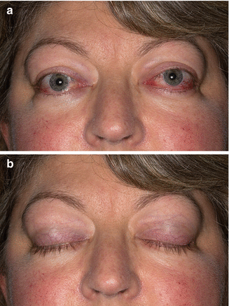
This patient was treated with block and replace regimen for her Graves’ disease. When reviewed in clinic 3 months later, she was complaining of pain around her eyes that becomes worse on eye movement. She has also noticed intermittent diplopia. Visual acuity was normal and her colour vision was intact. Figure 5.2 represents a picture of this lady.


Fig. 5.2
The patient 3 months after the initial presentation. (a) Conjunctival and eyelid abnormalities are clearly evident (see text). (b) Full eye closure rules out the possibility of exposure keratitis
Describe what you see?
The patient has periorbital odema, swollen and red eyelids, conjunctival injection and early chemosis on the left and swelling of the caruncle. She has full eye closure and therefore no risk of exposure keratitis.
What would you do at this stage?
This lady’s TED is showing clear signs of progression with a clinical activity score of 7/7. She has pain on eye movements, together with eyelid redness and swelling. She also has swelling of the caruncle, conjunctival redness and chemosis. Therefore, systemic treatment with steroid is advised. The lady was started on intravenous methylprednisolone using a EUGOGO-approved regimen. Alternatively, she can be treated with oral steroids, but these are generally less effective and associated with more systemic side effects [2, 3].
It should be noted that steroid use is a relative contraindication in our patient due to the diagnosis of diabetes. However, given the severity of the condition, methylprednisolone can be used with close monitoring of the blood glucose and adjustments in therapy as appropriate.
What would you need to monitor whilst on methylprednisolone therapy?
The patient should have baseline tests like any other patient starting on steroids (full blood count, U&Es, glucose, HbA1c and LFTs). Particular emphasis should be placed on liver function tests as methylprednisolone therapy can cause hepatitis and should be avoided in patients with a history of significant liver disease [4]. However, severe cases of hepatitis were generally related to the use of a particular regimen (1 g methylprednisolone daily for 3 days), which is perhaps best avoided. The authors prefer to use methylprednisolone at 500 mg doses weekly for 6 weeks, followed by 250 mg weekly for 6 weeks (cumulative dose of 4.5 g). Maximum dose of methylprednisolone used should not exceed 6.5 g, as per EUOGOGO guidelines.
What the best treatment strategy for the management of hyperthyroidism in TED?
There are no randomised controlled trials on this, which remains an area guided by personal experience rather than hard evidence. It is generally accepted that fluctuation of thyroid function should be avoided (particularly hypothyroidism), as this may exacerbate TED; therefore, the majority of these patients are treated with block and replace regimen during the active phase of TED [5].
Stay updated, free articles. Join our Telegram channel

Full access? Get Clinical Tree


