© Springer International Publishing Switzerland 2016
David S. Cooper and Cosimo Durante (eds.)Thyroid Cancer10.1007/978-3-319-22401-5_66. A Case of Multifocal Papillary Thyroid Microcarcinoma
(1)
Division of Endocrinology, Mayo Clinic Florida, Mayo Clinic College of Medicine, Rochester, Minnesota, USA
Case Presentation
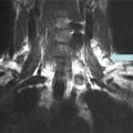
A 68-year-old female reported for evaluation of a 2 cm right thyroid nodule incidentally discovered on an MRI as part of an evaluation for shingles. Thyroid function tests are within normal limits. The patient had no history of radiation exposure involving the head and neck and no family history of thyroid cancer, but there was a family history of lymphocytic thyroiditis. The patient was asymptomatic and physical examination was unremarkable.
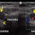
Neck ultrasound revealed a diffusely heterogeneous thyroid with a dominant 2.2 × 1.7 × 1.8 cm solid nodule in the right lower pole, a less well-defined 2.0 × 0.8 × 1.6 cm nodule in the right upper pole, a left lower pole 0.3 cm calcification without associated nodule, and a sub-centimeter isthmic nodule.
Fine-needle aspiration (FNA) of the two >1 cm nodules was performed with the cytology for the right lower pole nodule being suspicious for papillary thyroid cancer (PTC) (Bethesda class V).
Diagnosis/Assessment
The patient underwent a total thyroidectomy with central compartment neck dissection. The final pathology reported a 0.3 cm right lobe PTC and a 2.0 cm hyalinizing trabecular adenoma. In the left lobe, there were two foci of PTC measuring 0.2 and 0.4 cm. Margins were uninvolved by carcinoma. Background lymphocytic thyroiditis was present. All eleven lymph nodes excised were negative for tumor. The patient was staged T1aN0M0 (AJCC/UICC staging system, seventh edition) and radioactive iodine ablation of the thyroid bed was not recommended.
Postoperative Management
Following surgery the patient was placed on a less than fully suppressive dose of levothyroxine with a target serum thyroid-stimulating hormone (TSH) of 0.1–0.3 mU/L.
Outcome
A 24-month follow-up did not indicate any signs of recurrence by ultrasound. TSH-suppressed thyroglobulin (Tg) remained undetectable, with anti-Tg antibody titer declining from 156 IU/mL at 4 months to 59 IU/mL at two years postsurgery. Serum Tg by HPLC was undetectable.
Literature Review
Epidemiology
Thyroid cancer is the fastest-growing malignancy in incidence within and outside the United States with PTC representing over 85 % of cases [1]. This is at least in part attributable to tumors measuring less than 1 cm in diameter, most of which are diagnosed incidentally through increased use of diagnostic imaging, together with the widespread availability of high-resolution ultrasound that allows FNA of nodules as small as 3–5 mm in diameter.
The World Health Organization defines papillary thyroid microcarcinomas (PTMCs) as tumors measuring less than 1 cm in size [2]. These tumors currently represent about 50 % of all thyroid cancers [3], while their incidence in population, based on autopsy studies, varies between 5.3 and 35.6 % [4, 5].
Diagnosis
There are three common scenarios that can lead to the diagnosis of a PTMC: in the pathology specimen of a thyroid gland removed for benign disease (“incidental” PTMC), by surgery following FNA suspecting PTC, and by diagnosis of PTC in a cervical lymph node or distant metastasis (“occult” PTMC). A few decades ago, over 60 % of PTMCs were diagnosed either incidentally during thyroidectomy performed for benign etiologies or by open biopsy of a cervical lymph node. With the advances in ultrasound techniques and introduction of ultrasound-guided FNA, over the last three decades, almost half of PTMCs are diagnosed preoperatively [6].
An epidemiological review of two national registries reported that 26–33.5 % of PTMCs were multifocal [3], in concordance with other studies [7]; another study reported a frequency of multifocality as high as 42 % [8]. Up to 22 % of these tumors are found to be bilateral [9, 10]. Hay et al. reported multifocality in 20 % and bilaterality in 10 % of a series of 535 PTMCs [7]. The presence of multiple PTC foci in one lobe increases the likelihood that additional foci are present in the contralateral lobe, and by one study, multifocality is also increased in patients over age 45 [10].
Multiple studies have addressed the question whether the multiple foci of PTC arise independently of each other, as opposed to having a unifocal origin followed by intrathyroidal spread. These clonality analyses used various methods including assessment of RET/PTC rearrangement, X-chromosome inactivation, and analysis of BRAF mutation. Sugg et al. [11] reported that different synchronous tumor foci in the same thyroid gland exhibited different RET/PTC rearrangements in 15 (88 %) of a series of 17 patients, suggesting that most PTMCs arise in an independent fashion. These findings were later confirmed by Park et al. in a study of 61 cases [12]. Conversely, several other studies found that most multifocal PTMCs arise from intrathyroidal spread [13–16], while in other studies, a similar number of cases appeared to arise from independent foci and intrathyroidal spread [17, 18].
Risk Factors for Aggressive Disease
Several factors have been recognized as influencing the clinical outcome in PTC: the size of the tumor, age at diagnosis, and extrathyroid and extra-nodal extension. Certain histological subtypes such as tall cell, hobnail, and columnar cell variants of PTC are also considered to have a more aggressive behavior [19, 20]. PTMC is generally regarded as a low-risk disease and does not require aggressive treatment or intensive follow-up, with a disease-specific mortality rate less than 1 % [6]. Although the vast majority of thyroid microcarcinomas are low risk, with an excellent long-term outcome [19], a very small subset of these tumors (0.2–1 %) develop distant metastases and subsequent disease-related mortality. Clinical practice is in need of studies to identify biochemical and genetic markers of aggressiveness to assist in identification of this minority of patients, thereby facilitating individualized management [3, 7].
Risk factors for aggressive disease in PTMC have been extensively studied. They include male gender, multifocality [10], and extrathyroidal extension [8]. Hay [7] reported that N1 status at diagnosis increases the risk for locoregional recurrence to 18 % compared to 1 % in N0 patients, similar to findings reported by Wada in a series of 259 PTMCs [21].
Multifocality as a risk factor for recurrence in PTMC has been analyzed by several studies, with conflicting results. In a series of 900 patients with PTMC at the Mayo Clinic, Hay et al. found it as risk factor for recurrence [6]. Similarly, Malandrino et al. found multifocality to be more frequently associated with lymph node involvement, in addition to tumor size >6 mm and extrathyroidal invasion [3]. Conversely, Neuhold et al. did not find a difference with respect to lymph node involvement or increased risk of recurrence compared to unifocal PTMC [22].
Since multifocality is reported in 20–40 % of PTMCs, but these tumors have a significantly lower incidence of recurrence, additional factors must be considered to explain a more aggressive behavior in a small subset. In the recent decades, significant attention has been given to genetic features of PTCs, as predictors for tumor behavior. Among these, the V600E mutation in the BRAF gene has been linked to aggressive histopathologic features of PTC and was reported in a significant percentage of multifocal PTMCs [23]. The presence of V600E mutation in PTMC was found to be associated with higher clinical recurrence in low-risk PTMC, but not confirmed as an independent risk factor [9]. This discrepancy between the number of multifocal PTMCs displaying the V600E mutation and the risk of recurrence indicates that additional factors must contribute to the aggressive nature of some of these cancers. Niemeier et al. proposed a thyroid microcarcinoma predictive risk score comprised of presence of V600E mutation and several histopathological features including anatomic location, tumor size, tumor location with respect to the thyroid capsule, status of surgical margins, presence of infiltrative tumor border, tumor growth pattern, multifocality, extrathyroidal extension, degree of fibrosis, presence of psammoma bodies, and presence of lymphocytic thyroiditis [24]. This scoring system demonstrated 96 % sensitivity and 80 % specificity in differentiating more aggressive PTMC from tumors with less aggressive behavior [24]. Therefore, the management and follow-up of PTMC need to be individualized, taking into account other characteristics in addition to the tumor size alone.
Treatment of Multifocal PTMC
Once PTC is suspected by FNA, the recommended treatment consists of total or near-total thyroidectomy, potentially followed by radioactive iodine ablation of remnant thyroid tissue. These recommendations are endorsed by both the 2009 American Thyroid Association (ATA) guidelines and by the consensus report of the European Society of Endocrine Surgeons [25] and supported by the high incidence of multicentric tumors, with recurrence rates as high as 20 % in the remaining thyroid [26].
While for tumors >1 cm in diameter, total thyroidectomy is generally accepted as standard of care, in management of PTMC, the extent of surgery, the use of radioactive iodine, and TSH suppression are subjects of debate. The challenge consists in choosing the appropriate therapeutic measures so that recurrence-free survival and overall survival benefit exceeds the risks associated with extent of surgery (transient or permanent postoperative hypocalcemia and laryngeal recurrent nerve injury), RAI ablation (xerostomia, xerophtalmia, parotiditis), and prolonged iatrogenic hyperthyroidism (cardiac arrhythmias and loss of bone mass). For a PTMC confined to the thyroid and without lymph node extension, lobectomy is recommended in current ATA guidelines, if there is no definitive indication for removal of the contralateral lobe.
In the case of incidental microcarcinomas diagnosed following a thyroid lobectomy for a benign diagnosis, the lobectomy alone is reasonable in the absence of additional risk factors such as family history of thyroid cancer or a previous history of radiation to the head and neck [19]. Multifocality is associated with a slightly increased risk for structural disease recurrence; however, optimal surgical management is a matter of debate. In a large study on 535 consecutive cases of PTMC treated at the Mayo Clinic, Hay et al. concluded that PTMC has an excellent prognosis if the initial treatment consists of a total or subtotal thyroidectomy, while RAI did not improve the locoregional recurrence rate. The authors however recognize that “hemithyroidectomy as opposed to near-total or total thyroidectomy does not compromise survival,” but recommend an initial bilateral lobar resection (near-total thyroidectomy) in order to address the potential risk of cancer in the contralateral thyroid lobes [7]. Studies over the past decade focused on the outcome of patients with PTC who were treated with lobectomy and reported excellent recurrence-free survival rates [27]. A SEER database analysis of 23,605 patients with PTC reported that in low-risk patients, there was no association between lobectomy and poorer cause-specific or overall survival [28], findings confirmed by Mendelsohn et al. who concluded that in properly selected patients, lobectomy alone is associated with excellent disease-free survival [29].
Furthermore, there are proponents of observation rather than intervention in selected patients with unicentric PTMC. Ito et al. studied age as a prognostic factor in PTMC, in a cohort of 1235 cases, and found that in patients over 60 years old with subclinical, low-risk disease, observation with once or twice yearly ultrasonography is a reasonable option. In this study, regardless of age, multifocality was not found to be a significant risk factor for size enlargement, novel lymph node appearance, or risk of clinical disease.
Related posts:
 A Case of a Papillary Thyroid Cancer with Lymph Node Metastases Found on Prophylactic Central Neck Dissection (Subclinical Disease, Micrometastases)
Risks of Thyroid Hormone Suppression for Differentiated Thyroid Cancer in the Elderly
Radioiodine Therapy in Lactating Women with Higher-Risk Differentiated Thyroid Cancer
A Case of a Papillary Thyroid Cancer with Lymph Node Metastases Found on Prophylactic Central Neck Dissection (Subclinical Disease, Micrometastases)
Risks of Thyroid Hormone Suppression for Differentiated Thyroid Cancer in the Elderly
Radioiodine Therapy in Lactating Women with Higher-Risk Differentiated Thyroid Cancer
 A Patient with a Large Hürthle Cell Carcinoma of the Thyroid and Nodal Metastases
 Papillary Thyroid Carcinoma Diagnosed During Pregnancy
 Timing and Extent of Surgery for a Pediatric Patient with Hereditary MTC and Positive Screening for the S891A RET Mutation
Stay updated, free articles. Join our Telegram channel

Full access? Get Clinical Tree


