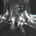
Fig. 30.1
Upper left: non-contrast-enhanced CT showing right paratracheal mass (white arrow). Lower left: 18FDG-PET-CT showing right paratracheal recurrent tumor (white arrow). Upper right: 18FDG uptake with a standard uptake value of 10.5. Lower right: 18FDG-PET showing uptake in three lesions on the right side of the neck—from top to bottom—level II, paratracheal and pretracheal, shown by black arrows
Due to the size and progressive nature of the tumor in the neck, reoperative surgery in the neck and upper mediastinum was proposed by the multidisciplinary tumor board. Preoperative laryngoscopy was normal. Surgery was performed using intermittent recurrent nerve neuromonitoring. Intraoperative findings revealed that the level II metastasis invaded the posterior aspect of the internal jugular vein which was sacrificed. The right paratracheal node completely surrounded the inferior laryngeal (recurrent) nerve and superficially invaded the esophageal muscularis, which was resected with a shaving technique, with no perforation of the esophageal mucosa. The nerve was resected en bloc with the tumor mass. The pretracheal node was also resected en bloc with the paratracheal mass. Final pathology revealed three lymph node metastases (20–25 mm each) with the tall cell variant of papillary thyroid carcinoma and extranodal extension. The postoperative course was uneventful, with a satisfactory vocal outcome despite resection of the recurrent nerve.
Assessment and Literature Review
Evaluation and Diagnosis
How Do We Know That This Patient Has RAI-Refractory Disease?
After the fourth administration of a therapeutic dose of RAI, no uptake was seen on SPECT imaging, but the micrometastases were still visible on CT and Tg was still elevated, proof of persistent disease despite RAI [1].
Was FNAC Necessary if 18FDG-PET-CT Showed Significant Uptake?
The reported specificity of 18 FDG-PET-CT for recurrence in the neck is 67 % [2], whereas the specificity of ultrasound is 89 %. Ultrasound-guided FNAC with Tg measured on the needle aspirate fluid has a reported specificity of 95 % [3]. Thus, FNAC with measurement of Tg in the needle washout fluid, when possible, improves diagnostic accuracy and avoids the pitfall of false-positive readings of18FDG-PET-CT. FNAC also rules out other tumors, such as lung or head and neck cancer in this older patient with a history of smoking. FNAC is indicated for diagnostic purposes only when a specific treatment can be proposed. Small (<8 mm) suspicious lymph nodes may be followed safely in many cases, with little or no increase in size over time or regional complications. Surgery may be suggested for larger metastases, however, to avoid regional complications in case of tumor progression and to optimize disease-free survival.
What Does the Previous Course of the Disease Tell Us About Prognosis?
The extrathyroidal extension of the disease (pT3), the initial stage of the neck disease (N1b), the age of the patient at first diagnosis (59 years old), and his gender are risk factors for lower disease-free survival [4, 5]. The aggressive histopathological variant of differentiated thyroid carcinoma (tall cell) has also been shown to be a risk factor for recurrent/persistent disease and worse survival [6, 7]. In rare cases, tall cell carcinoma (> or =50 % tall cells) and carcinoma with tall cell features (30–49 % tall cells) have also been found to show anaplastic transformation in the recurrent tumor [6].
The lymph node ratio (the ratio of the number of metastatic nodes found to the total number of resected lymph nodes)—13/20 or 0.65 in this patient after the first reoperation—has also been shown to portend a lower rate of disease-free survival. A rate of 0.3 or above was associated with a 3.4 times higher rate of persistent or recurrent disease, in the study by Vas Nunes et al. [8]. A cutoff of 0.7 was associated with worse prognosis for Schneider et al. [9]. A number of metastatic nodes exceeding ten have also been shown to be a risk factor for persistent/recurrent disease for papillary thyroid carcinoma [10]. Extranodal spread of disease—a further risk factor [10]—was not noted on the pathological report from the first reoperation in the neck in our patient, although it was evident after the operation at our institution. Finally, the presence of macroscopic (>1 cm) metastatic nodes resected at the first operation has been shown to be a factor for persistent/recurrent neck disease [11]. Unfortunately, we were not able to verify the size of the metastatic nodes resected at the first operation in our patient, this operation having been performed at an outside institution.
What Other Imaging or Explorations Could or Should Be Performed to Determine Resectability of the Neck Disease?
In a reoperative situation with macroscopic surgical targets in proximity to the carotid artery, trachea, and esophagus, morphological imaging such as contrast-enhanced CT or MRI may improve prediction of local invasion and aid surgical planning [12]. With recurrent disease close to the esophagus, endoscopic ultrasound may also be helpful in determining the depth of any invasion of the esophageal muscle wall [13]. Finally, laryngoscopy is highly recommended in cases with recurrent disease before reoperation, to detect recurrent nerve paralysis from a previous operation or from locally invasive cancer [14, 15]. Preoperative laryngeal mobility was normal for our patient.
Management
What Are the Treatment Options?
Due to the slowly progressive nature of the disease and the relatively good prognosis over the short to medium term, surgery is generally recommended for potentially resectable lesions, unless undue morbidity is to be expected [16, 17]. External beam radiation therapy to the neck is an option for progressive but unresectable disease [17–19]. Other local therapies such as alcohol injection, radiofrequency ablation, or cryotherapy have not been widely studied in the treatment of large (>1 cm) neck recurrences. Systemic therapy with small molecule tyrosine kinase inhibitors is generally reserved for progressive distant metastases of significant volume, due to the long-term nature of this treatment and its toxicities. Confirmed invasion of the viscera or brain may be a contraindication to this type of systemic therapy with anti-angiogenic activity, however, due to the risk of perforation, fistula, or hemorrhage [20].
What Extent of Surgery Is Recommended?
For our patient, several factors favored surgery to remove the neck metastases: the good general health of the patient despite his comorbidities and age, the size of the metastases >1 cm, RAI-refractory disease, the progression of serum Tg, resectability ascertained on contrast-enhanced CT, the small volume of the lung metastases and their stability over 7 years, and the absence of other detectable lesions. The aim of surgery was to reduce tumor burden and to reduce the risk of symptoms in the neck from vascular and visceral invasion over time.
Why Was the Recurrent Nerve Resected if It Was Functional?
Dissection of the tumor off of the nerve sheath is technically feasible, but generally leaves small disease on the nerve, with resection classified as R1 (microscopic residual disease) or R2 (macroscopic residual disease) [21]. In our patient, however, the distal end of the nerve was entirely encased by the tumor, and nerve preservation would have required leaving macroscopic residual tumor near the larynx, trachea, and esophagus (R2 resection). Our aim was to optimize locoregional control in the neck and minimize the risk of invasion of the trachea and/or esophagus. With RAI-refractory tumors, this goal can be attained only with a macroscopically complete surgical resection.
Swallowing may be affected postoperatively if there is recurrent nerve damage, with aspiration of liquids, and patients should be monitored and receive swallowing rehabilitation if necessary. Voice outcomes are variable after resection of the recurrent nerve due to several factors: compensation from the contralateral side, bilateral innervation of the interarytenoid muscle, variable muscle atrophy, and some degree of reinnervation from nerve anastomoses and from the regional autonomous nerve system. Voice can be surgically improved with several methods based on volume augmentation of the paralyzed vocal fold or medialization of the vocal fold or the arytenoid cartilage, generally with excellent results [22]. Our patient did not have any postoperative dysphagia and considered his voice satisfactory; he declined any type of intervention to improve his voice.
What Outcome Can Be Expected?
A survival rate of 10 % at 10 years and 6 % at 15 years has been reported for patients harboring RAI-refractory metastatic disease [23]. In the same study, however, an intermediate group of patients aged >40 years with RAI-refractory disease but with low tumor burden and micronodular lung metastases had a survival rate of 67 % at 10 years. Our patient falls into this intermediate group with a relatively good prognosis (better than many other solid tumors). This intermediate prognosis justifies locoregional treatments when possible to maintain a low tumor burden and avoid symptoms from progressive disease. For patients with multiple recurrences in the neck, Kim et al. [24] showed a 10-year disease-specific survival rate of 83 % as compared to patients with no recurrence or only 1 recurrence whose 10-year disease-specific survival rate was 100 %.
Reoperative surgery in the neck must be preceded by a meticulous preoperative imaging and planning, in order to foresee possible complications or sequelae and inform the patient preoperatively but also to find and completely map all of the neck metastases in order to resect all of the disease at the time of the operation [25, 26]. Morbidity for reoperation is higher than for primary neck dissection, with a higher risk of unintended nerve injury (recurrent, spinal accessory, and phrenic nerves in particular), tracheal or esophageal injury, and chyle leak from thoracic duct injury [26–28]. Intentional resection of the recurrent nerve, more frequent in the reoperative setting [25], should be discussed with the patient preoperatively, with information regarding the effectiveness of a vocal rehabilitation procedure secondarily if needed.
Recurrence after reoperative surgery has been reported to occur in 40–66 % of cases [26, 27, 29–31]. When evaluated, re-recurrence may be related to inherent disease aggressiveness (particularly after an apparently disease-free interval), but insufficient preoperative workup, “missing” metastatic nodes at the first reoperation, can also be a cause of recurrent/persistent disease [26]. A persistently elevated stimulated Tg level after reoperation is a risk factor for further structural recurrence/persistence [29].
Several techniques have been developed to aid in localizing recurrences, which are often difficult to locate due to scarring from previous surgery. Intraoperative ultrasound may be performed, but requires training in ultrasonography or bringing the radiologist into the operating room. Radio-guided surgery with RAI (when uptake is present) may be helpful, particularly for metastatic nodes in the mediastinum or retropharyngeal nodes, not seen on ultrasound [32]. 18 FDG-PET -guided surgery has been described but is not yet widely employed due to technical constraints [33, 34]. Harpoon-guided (hooked-needle) surgery has been described, but may be hazardous for small metastases close to major vascular structures such as the carotid artery and internal jugular vein [35]. Ultrasound tattoo-guided surgery, using colloidal charcoal or methylene blue, is relatively simple to implement, with high rates of localization, but is only applicable to lesions that can be visualized on ultrasound and that are accessible with a needle [36–38]. Due to the size of the recurrences which were well visualized on CT, and their anatomic locations, we did not use localizing techniques in this patient, however.
Related posts:
 A Case of Multifocal Papillary Thyroid Microcarcinoma
A Patient with Papillary Thyroid Cancer and Biochemical Evidence of Disease at the One-Year Follow-Up Visit
A Case of Papillary Thyroid Cancer Without Aggressive Histological Features with Nodal Metastases in an Older Patient
A Case of Multifocal Papillary Thyroid Microcarcinoma
A Patient with Papillary Thyroid Cancer and Biochemical Evidence of Disease at the One-Year Follow-Up Visit
A Case of Papillary Thyroid Cancer Without Aggressive Histological Features with Nodal Metastases in an Older Patient
 A Patient with a Large Hürthle Cell Carcinoma of the Thyroid and Nodal Metastases
 Papillary Thyroid Carcinoma Diagnosed During Pregnancy
 Timing and Extent of Surgery for a Pediatric Patient with Hereditary MTC and Positive Screening for the S891A RET Mutation
Stay updated, free articles. Join our Telegram channel

Full access? Get Clinical Tree


