© Springer International Publishing Switzerland 2016
David S. Cooper and Cosimo Durante (eds.)Thyroid Cancer10.1007/978-3-319-22401-5_88. A Case of a Papillary Thyroid Cancer with Lymph Node Metastases Found on Prophylactic Central Neck Dissection (Subclinical Disease, Micrometastases)
(1)
Division of Endocrinology and Internal Medicine, Mayo Clinic and College of Medicine, 200 First Street SW, Rochester, MN 55905, USA
Keywords
Papillary thyroid carcinomaTotal thyroidectomyUnilateral lobectomyCentral compartment dissectionMinimal extrathyroid extensionRadioiodine remnant ablationPrognostic scoringTNM stagingThyroxine-suppressive therapySerum thyrotropinSerum thyroglobulinNeck ultrasoundAbbreviations
TSH
Thyroid stimulating hormone thyrotropin
US
Ultrasound
PTC
Papillary thyroid carcinoma
cTNM
Clinical tumor node metastasis
pTNM
Postoperative tumor node metastasis
AJCC
American Joint Committee on Cancer
UICC
International Union Against Cancer
CSM
Cause-specific mortality
AGES
Age grade extent size
MACIS
Metastases age completeness invasion size
AMES
Age metastases extrathyroid size
NNM
Neck nodal metastases
TST
Thyroxine-suppressive therapy
Tg
Thyroglobulin
RAI
Radioactive iodine
RRA
Radioiodine remnant ablation
WBS
Whole body scan
CT
Computerized tomographic
OPTC
Occult papillary thyroid carcinoma
DM
Distant metastases
BLR
Bilateral lobar resection
RNR
Regional neck recurrence
ATA
American Thyroid Association
ETA
European Thyroid Association
FDA
Food and Drug Administration
BST
Bilateral subtotal thyroidectomy
UL
Unilateral lobectomy
NTT
Near-total thyroidectomy
TT
Total thyroidectomy
LRR
Locoregional recurrence rate
R
Recommendation
MSKCC
Memorial Sloan Kettering Cancer Center
NCDB
National cancer data base
SEER
Surveillance epidemiology, and end results
Case Discussion
Presentation
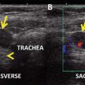
In January 2008, a 24-year-old woman saw her gynecologic nurse practitioner, who found an isthmic thyroid nodule. The patient was asymptomatic. She had no personal or family history of thyroid disease and had not been exposed in earlier life to head or neck irradiation. Her serum TSH was 2.1 mIU/L. A neck ultrasound (US) revealed a normal-sized thyroid gland and at the level of the isthmus just to the right of midline, a 9 mm isoechoic, solid nodule, correlating with the palpable finding. This nodule was avascular on color Doppler imaging and had no suspicious sonographic features. She was referred to an endocrinologist (the author), who felt that a guided biopsy was not indicated, but recommended a recheck neck US examination in 6 months.
Diagnosis
In August 2008, a repeat US showed the nodule to be slightly larger, now measuring 1.1 × 1.5 × 0.8 cm. An US-guided biopsy was performed; smears were positive for malignancy, and the cytologic features were consistent with papillary thyroid carcinoma (PTC). Chest X-ray was negative; vocal cord check showed normal motion bilaterally.
Surgical Management
After obtaining the diagnostic biopsy, the author discussed the expected outcome of patients presenting with a so-called “occult” PTC, as defined by Woolner et al. as a PTC with maximal tumor dimension of “1.5 cm or less” [1, 2]. The author recommended surgery, rather than observation, and advised a timely consultation with an experienced endocrine surgeon [3]. She noted that the neck US did not show any suspicious cervical adenopathy, but recommended for the patient a “near or total thyroidectomy with central compartment lymphadenectomy” [4–7]. She discussed the potential risks of the operation [8] and the need for lifelong thyroid hormone supplementation.
In August 2008, the patient underwent a “total thyroidectomy with central compartment lymphadenectomy.” All removed specimens were submitted to immediate examination of frozen sections and the results conveyed intraoperatively to the surgeon in the operating room [9]. The final histologic report showed the primary tumor to be a histologic Broders [10–12] grade 1 (of 4) classical PTC, measuring 1.3 × 1.0 × 0.8 cm, and situated in the isthmus. The tumor focally extended into the peri-thyroidal soft tissue [13, 14], but the surgical margins were free of involvement. The contralateral lobe contained only benign thyroid tissue. Microscopic examination of the central compartment level VI dissection specimen revealed metastatic PTC in multiple (3 of 3) central compartment level VI nodes. Contemporary pTNM staging, according to the AJCC/UICC sixth edition [15], was stage I (pT3 N1A MO). The case would be classified as “low risk for cause-specific mortality” (CSM) because of an AGES score of 0.26 and a MACIS score of 3.47 but also by the AMES prognostic classification system [11, 16, 17].
Assessment During First Postoperative Year
The patient, despite the presence of microscopic extrathyroidal extension and central compartment neck nodal metastases (NNM), was classified on postoperative day 1 as having a PTC at minimal risk of CSM. With a MACIS score <6, as per our institutional practice since 1994 [18–21], she was advised to leave the hospital on thyroxine-suppressive therapy (TST) and return in 6–8 weeks for a neck ultrasound and a recheck of serum TSH and serum thyroglobulin (Tg). She was not advised to have radioiodine remnant ablation (RRA) nor was she given a recommendation for a postoperative whole body scan (WBS) with radioactive 123I.
When she returned in October 2008, her serum TSH was 0.1 mIU/L, her Tg antibody screen was negative, and her serum Tg was 0.2 ng/ml. A neck US showed no evidence of thyroid bed nodules or suspicious neck adenopathy. The patient was advised to continue with her TST, return for review at one postoperative year with repeat studies, including a neck US as well as serum TSH and Tg measurements. She was told that her prognosis should be excellent [10–12] and that there was probably only a 5–10 % chance [4, 6, 18, 20] in future years of finding any further residual microscopic cervical lymphadenopathy. She was also advised that, if such cervical nodal metastatic disease was to be discovered, she would likely be treated with US-guided percutaneous ethanol ablation at the Mayo Clinic [22–24]. At 14 postoperative months, she returned for follow-up. Her neck US was again negative for locoregional recurrence; TSH was 0.08 mIU/L and serum Tg was again 0.2 ng/mL.
Surveillance from One Through Six Postoperative Years
For the next 5 years (2010–2014), the patient was followed annually. Serum TSH levels on TST during 2010 through 2013 varied between 0.1 and 0.3 mIU/L; Tg levels were on two visits, 0.1, and on three visits, 0.2 ng/mL. On the last review visit in November 2014 (at 74 postoperative months), the serum TSH was in the lower part of the normal range at 0.4 mIU/L, the serum Tg was stable at 0.2 ng/mL (<0.1 undetectable), and the neck US was negative for recurrence.
Literature Review
Postoperative Outcome in Woolner’s “Occult” PTC
At the 1959 American Goiter Association meeting, Dr. LB Woolner drew attention to the increasing prevalence of small, possibly biologically unimportant, papillary carcinomas. Most, he found, were 3 mm–1 cm in diameter, but he considered that “an arbitrary upper limit of size of 1.5 cm in greatest dimension” was appropriate to describe this increasingly recognized group. In 1960, he defined OPTC [1] as a “papillary carcinoma 1.5 cm or less in diameter with or without lymph node metastases.” In his earliest study of 140 such patients treated during 1920–1965, he reported that none developed distant metastases (DM) or died from OPTC [1]. In a later study published in 1980, he followed 137 cases for a mean of 25 years and was only able to define one possible death from OPTC, but he did note that the “exact cause of death remained in question” [2]. His 1980 study concluded that “radical surgery or medical extirpation of all thyroid tissue is unnecessary in the treatment of this (non-lethal and curable) disease” [2].
In a 1986 study of 859 PTC patients treated during 1946 through 1970, McConahey and colleagues identified 396 OPTC (46 %), and they noted that the risk of CSM increased progressively as the size of the tumors increased [10]. The CSM rate per 1000 cases was 0.8 for OPTC, 3.8 for tumors 1.6–3.9 cm diameter, and 12.6 for those 4 cm or larger (p < 0.0001). In this study, four fatal cases of OPTC were identified. All four were middle aged or older men (47–70 years of age at diagnosis); one patient had a grade 2 tumor and two had distant metastases at the time of initial presentation.
In 2002, our group reported the postoperative course of 1205 OPTC patients consecutively treated during 1940–2000, over a median follow-up period of 14 years [25]. Bilateral lobar resection (BLR) was the preferred surgical procedure in 89 %. Neck nodes were resected in 52 % of cases; RRA was given within six postoperative months in 27 %. Of 1190 patients having potentially curative surgery (no distant metastases and complete primary tumor resection), five developed postoperative distant metastases and five died of OPTC (20-year CSM rate of only 0.4 %). Overall, 20-year rates for local and regional recurrences were 2 % and 7 %, respectively, and were higher after lobectomy than BLR (p < 0.001). After BLR, the risks of local recurrence or distant spread were not decreased by RRA. When regional nodal recurrence (RNR) was studied after BLR in 367 node-positive patients, the 20-year rate of 15 % after RRA and BLR was insignificantly different (p = 0.23) from the 13 % seen after only BLR. For the 693 node-negative patients treated by BLR alone, the 20-year RNR rate of 1 % was not improved by RRA. From these results, we concluded that routine RRA, after BLR with complete tumor excision, was no longer justifiable in the management of patients presenting with small (<16-mm diameter) PTC.
Related posts:
 A Case of Multifocal Papillary Thyroid Microcarcinoma
Risks of Thyroid Hormone Suppression for Differentiated Thyroid Cancer in the Elderly
Radioiodine Therapy in Lactating Women with Higher-Risk Differentiated Thyroid Cancer
A Case of Multifocal Papillary Thyroid Microcarcinoma
Risks of Thyroid Hormone Suppression for Differentiated Thyroid Cancer in the Elderly
Radioiodine Therapy in Lactating Women with Higher-Risk Differentiated Thyroid Cancer
 A Patient with a Large Hürthle Cell Carcinoma of the Thyroid and Nodal Metastases
 Papillary Thyroid Carcinoma Diagnosed During Pregnancy
 Timing and Extent of Surgery for a Pediatric Patient with Hereditary MTC and Positive Screening for the S891A RET Mutation
Stay updated, free articles. Join our Telegram channel

Full access? Get Clinical Tree


