Quality indicator
Grade of recommendation
Appropriate indication
1C +
Informed consent is obtained, including specific discussion of risks associated with colonoscopy
3
Use of recommended postpolypectomy and post-cancer resection surveillance intervals
1A
Use of recommended ulcerative colitis/Crohn’s disease surveillance intervals
2C
Documentation in the procedure note of the quality of the preparation
2C
Cecal intubation rates (visualization of the cecum by notation of landmarks and photo documentation of landmarks should be present in every procedure)
1C
Detection of adenomas in asymptomatic individuals (screening)
1C
Withdrawal time: mean withdrawal time should be ≥ 6 min in colonoscopies with normal results performed in patients with intact anatomy
2C
Biopsy specimens obtained in patients with chronic diarrhea
2C
Number and distribution of biopsy samples in ulcerative colitis and Crohn’s colitis surveillance. Goal: 4 per 10-cm section of involved colon or approximately 32 specimens per case of pancolitis
1C
Mucosally based pedunculated polyps and sessile polyps < 2 cm in size should be endoscopically resected or documentation of unresectability obtained
3
Incidence of perforation by procedure type (all indications vs. screening) is measured
2C
Incidence of postpolypectomy bleeding is measured
2C
Postpolypectomy bleeding managed nonoperatively
1C
Interval Cancers and Deaths and Surrogate Measures
The primary goal of colonoscopy in most instances is the prevention of colorectal cancer-related death [8]. Results of the National Polyp Study in 1993 showed that colonoscopy reduced the incidence of colorectal cancers by 76–90 % [9]. In a case–control study using Surveillance, Epidemiology and End Results (SEER) datasets, Baxter et al. demonstrated a 60 % reduction in death from colorectal cancer. This protective association was strongest when colonoscopies were performed by gastroenterologists for distal lesions [10]. Despite the advances in colonoscope technology, the gastroenterologic societies realized that the protective benefits of colonoscopy were not as high as previously thought and that several explanations may account for these findings.
“Interval cancers,” colorectal cancers found at or before the next recommended screening/surveillance colonoscopy, are thought to be partly explained by missed lesions during the index colonoscopy [8, 11–13]. Large, retrospective, population studies from the USA and Canada have been performed to estimate the prevalence of interval cancers [11]. Findings were impressive, with 7.2–9 % of colorectal cancers meeting definition of interval cancers [13–15] (Table 7.2). There was a consistently higher probability of interval cancer occurring in the proximal compared to the distal colon.
Table 7.2
Frequency and location of interval CRCs. (Adapted from Ref. 51. With permission from Elsevier)
Study | Data source | Total detected cancers, n | Interval cancers | ||
|---|---|---|---|---|---|
Overall, n (%) | Proximal, n (%) | Distal, n (%) | |||
Baxter et al. 2011 [13] | Ontario cancer registry (2000–2005) | 24,213 | 1260 (9 %) | 676 (12.4 %) | 584 (6.8 %) |
Manitoba cancer registry (1992–2008) | 4883 | 388 (7.9 %) | 225 (11.3 %) | 147 (5.3 %) | |
Cooper et al. 2012 [15] | SEER-medicare database (1994–2005) | 57,839 | 4192 (7.2 %) | 2851 (9.9 %) | 1253 (4.5 %) |
While it is proposed that some interval cancers may arise from neoplastic lesions that harbor genetic features that are associated with a more rapid progression to cancer, as well as lesions that may have been incompletely resected, there is strong evidence to demonstrate that the quality of the colonoscopy is related to the rate of interval cancers [11, 12, 17–19].
The adenoma detection rate (ADR) , which is the proportion of average-risk patients undergoing screening colonoscopy in whom an adenoma or colorectal cancer is found, is regarded as a robust measure of colonoscopy performance quality that correlates with subsequent cancer risk [20–22]. It has been suggested that adenomas should be detected in 25 % or more of men and 15 % or more of women 50 years of age and older [23]. However, numerous studies have been published showing significant heterogeneity in endoscopist ADR. In a provocative landmark study by Kaminski et al., in which 45,026 patients involved in a Polish nationwide colorectal cancer screening program were followed over time, and interval cancers were determined at the scheduled time of surveillance colonoscopy, endoscopists with ADRs less than 20 % (categorized as less than 11.0 %, 11.0–14.9 %, 15.0–19.9 %, and 20.0 % or more) had a more than tenfold higher rate of interval colorectal cancers than those with higher ADRs [22]. Another more recent study that also supports the relationship between ADR and interval cancer comes from Corley and colleagues. Physician ADR (range: < 20.3 %– ≥ 32.0 %) was found to be an independent predictor of subsequent colorectal cancer risk following a negative colonoscopy, findings that were consistent for proximal and distal cancers, and irrespective of patient sex [24]. What remains to be determined is at what threshold we start to observe no further protective benefit of a very high ADR.
Some interval cancers may arise from rapidly growing lesions that develop de novo between scheduled colonoscopies. It is now well recognized that some colorectal cancers arise from a different carcinogenesis pathway, arising from the sessile serrated adenoma pathway. This pathway is characterized by mutations in the BRAF oncogene, gene promoter hypermethylation (i.e., CpG island methylator phenotype; CIMP) and a more rapid progression to colorectal cancer; these lesions are also more prevalent in the proximal colon, a location where we recognize colonoscopy to be less protective for colorectal cancer compared to the distal colon [25–27]. Recent studies suggest that these sessile serrated adenomas may partially account for some of the interval cancers . However, in a retrospective study by Kahi and colleagues, the proportion of screening colonoscopies with at least one proximal serrated polyp was 13 %, and, furthermore, endoscopist ADR correlated strongly with proximal serrated polyp detection rates [27]. To date, ADR remains the best surrogate for quality in colonoscopy and efforts are being undertaken to improve physician ADRs across many institutions.
Withdrawal time, the time measured from when the colonoscope reaches the cecum to the time the scope is withdrawn from the anus in the absence of polyp removal, has also been studied as a quality metric in colonoscopy. Studies have demonstrated that a withdrawal time of ≥ 6 min or more increased the detection of neoplastic lesions during colonoscopy in patients with intact colons [8]. However, due to heterogeneity in patient anatomy and lengths of colons, application of this standard is not feasible for every case [8].
Clinical studies have shown mixed results on improvements in adenoma detection with implementation of a longer withdrawal time [21]. Studies that have evaluated total withdrawal time alone as well as with feedback on performance failed to show statistically significant improvements in adenoma detection, while some showed improvement in nonadenomatous polyp detection [28–35]. When multiple interventions were implemented in studies by Imperiali et al. and Shaukat et al., including a 1 % financial penalty for those endoscopists who did not achieve a ≥ 6-min withdrawal time for > 95 % of examinations in the latter study, no statistically significant changes in ADR were observed [36, 37]. However, in a study by Barclay and colleagues, in which an audible timer was used during withdrawal (implementing an 8-min withdrawal time) in addition to enhanced inspection techniques, ADR increased by 50 % compared to baseline, a statistically significant finding (P < 0.0001) [21, 38]. In summary, mandating longer withdrawal time alone is not likely to increase the rate of adenoma detection and ultimately reduce the incidence and mortality of colorectal cancer [8].
Cecal intubation, defined as the process where the colonoscope reaches a point proximal to the ileocecal valve with complete visualization of the entire cecum, should be achieved in ≥ 90 % of all colonoscopies and in ≥ 95 % of cases for screening colonoscopies. Documentation of reaching this landmark should be confirmed with photography of the cecal landmarks (i.e., appendiceal orifice and ileocecal valve) [8]. This quality indicator has been proposed due to the well-known findings that a large portion of colorectal neoplasms is located in the proximal colon, including the cecum [8].
The quality of colonoscopy is also assessed by process measures for health-care delivery [6]. Documentation of various measures has been proposed by the ASGE as well as the ASGE/ACG Task Force on Quality in Endoscopy [8, 39]. Moving forward with these proposed recommendations, the Quality Assurance Task Group of the National Colorectal Cancer Roundtable (NCCRT) developed a standardized colonoscopy reporting and data system (CO-RADS) to improve the quality of colonoscopy [40]. Procedure reports should be created by programs to allow systematic documentation of the details of the colonoscopy that would include the indication(s), anatomic extent of the examination, findings, and complications, among others. The complete list of the recommended elements [39] is as follows:
1.
Date of procedure
2.
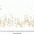
Patient identification data
3.
Endoscopist(s)
4.
Assistant(s)
5.
Documentation of relevant patient history and physical examination
6.
Indication of informed consent
7.
Endoscopic procedure
8.
Indication(s)
9.
Type of endoscopic instrument
10.
Medication (anesthesia, analgesia, sedation)
11.
Anatomic extent of examination
12.
Limitation(s) of examination
13.
Tissue or fluid samples obtained
14.
Findings
15.
Diagnostic impression
16.
Results of therapeutic intervention (if any)
17.
 Toward a Learning Health-Care System: Use of Colorectal Cancer Quality Measures for Physician Evaluation
Medical Legal Aspects of Quality Improvement
Areas for Future Research
Toward a Learning Health-Care System: Use of Colorectal Cancer Quality Measures for Physician Evaluation
Medical Legal Aspects of Quality Improvement
Areas for Future Research
 Quality Indicators and Benchmarks for Guideline-Recommended Fecal Occult Blood Tests
History and Overview of the National Quality Strategy
Quality Indicators and Benchmarks for Guideline-Recommended Fecal Occult Blood Tests
History and Overview of the National Quality Strategy
 Quality Indicators for CT Colonography


Complications (if any)
Related posts:
Toward a Learning Health-Care System: Use of Colorectal Cancer Quality Measures for Physician Evaluation
Medical Legal Aspects of Quality Improvement
Areas for Future Research
Quality Indicators and Benchmarks for Guideline-Recommended Fecal Occult Blood Tests
History and Overview of the National Quality Strategy
Quality Indicators for CT Colonography
Stay updated, free articles. Join our Telegram channel
Full access? Get Clinical Tree
