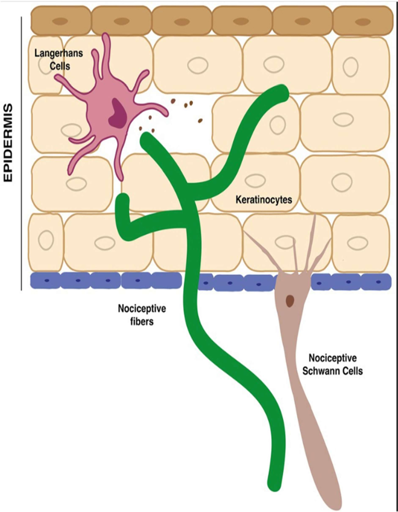
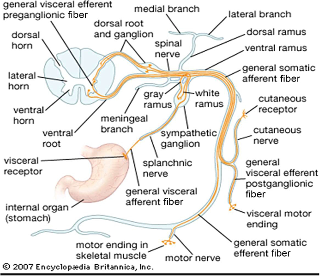
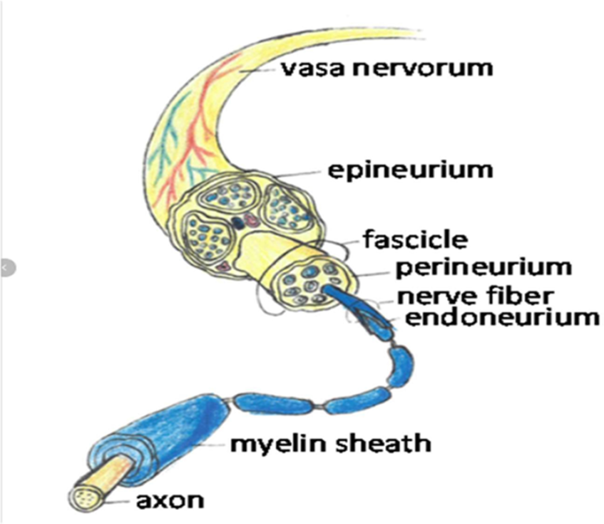
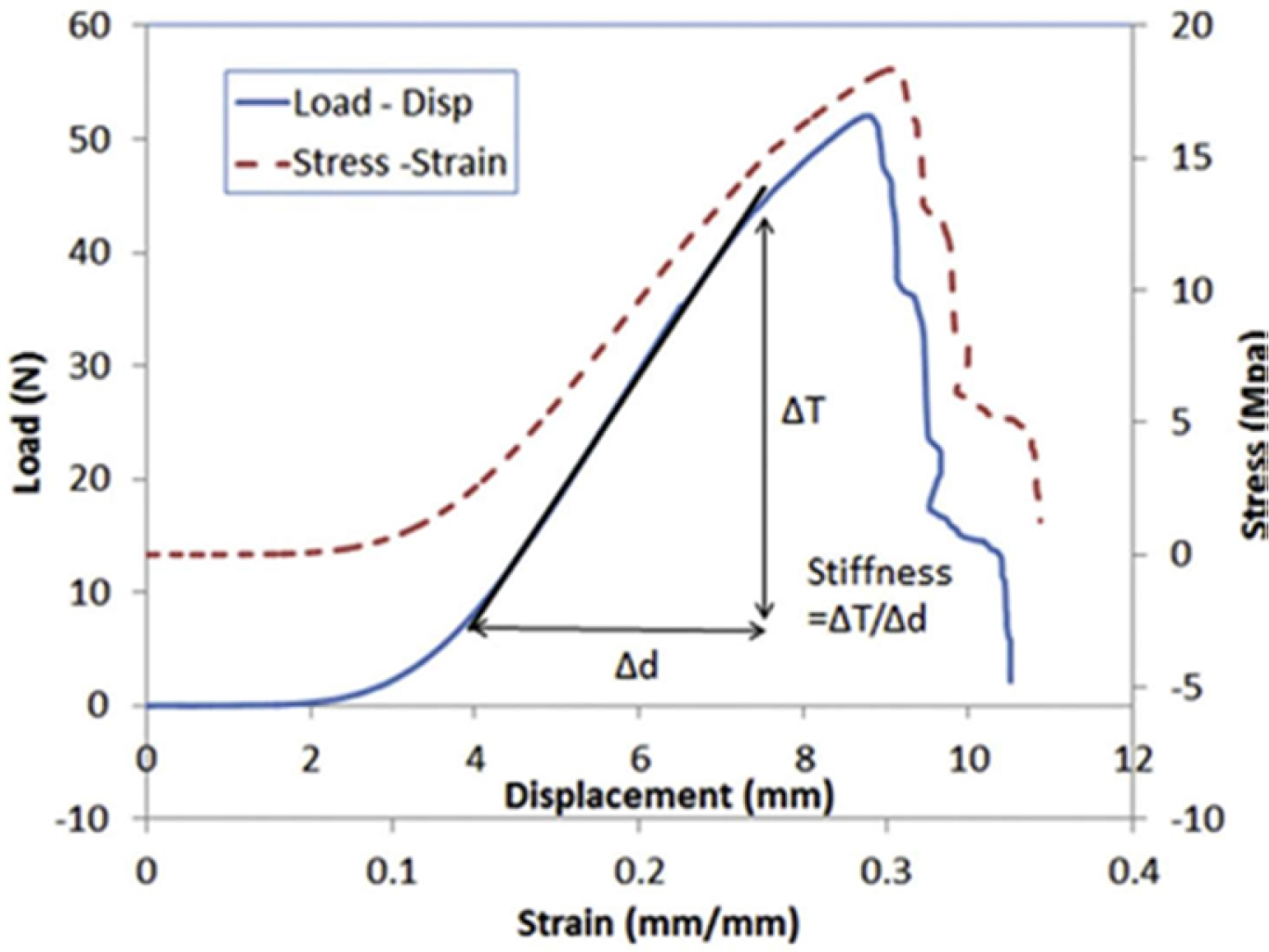
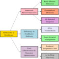
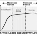
Physiotherapy plays a pivotal role in the rehabilitation and restoration of functional abilities following injury, illness or chronic health conditions. This chapter explores the multidimensional approach of physiotherapy, emphasizing its importance in improving mobility, relieving pain and enhancing the quality of life across diverse patient populations. By integrating manual therapy, exercise prescription, electrotherapy and patient education, physiotherapists address musculoskeletal, neurological and cardiopulmonary challenges with tailored treatment plans. This chapter highlights the evolving landscape of physiotherapy practice, from traditional hands-on techniques to advanced modalities such as dry needling, kinesio taping and tele-rehabilitation. A thorough understanding of patient assessment and evidence-based interventions ensures optimal outcomes, especially in cases involving post-operative care, sports injuries and chronic conditions such as arthritis and stroke. Furthermore, the collaborative nature of physiotherapy in multidisciplinary healthcare teams fosters holistic patient recovery. Emphasis is also placed on patient motivation, adherence to therapy and psychological support as integral to achieving therapeutic goals. Recent research underscores the preventative role of physiotherapy in minimizing the recurrence of injuries and managing long-term disability. This chapter aims to present a comprehensive overview of physiotherapy as a dynamic and essential healthcare discipline, advocating for its broader integration in both clinical and community settings. Through real-world case references and clinical insights, the study reinforces physiotherapy’s impact on improving functional independence and promoting long-term wellness. Figure 5.1. Cellular and neural components of the epidermis involved in nociception. The nervous system is divided into central and peripheral, where central is further divided into brain and spinal cord and the peripheral nervous system is divided into the autonomic nervous system and somatosensory nervous system. This chapter covers the somatosensory nervous system. The somatosensory nervous system is a combination of peripheral nerves and associated structures. This system senses stimuli through an afferent pathway and controls the response via voluntarily activated muscles in an efferent pathway (Pacifico et al. 2023). Figure 5.1 shows cellular and neural components of the epidermis involved in nociception. Nerves and nerve roots are mechanically and physiologically distinct from each other. While a nerve contains two different types of neurons, the nerve root contains only one specific type of neuron specific to its function. The different types of neurons are motor and sensory. Injury to a single nerve has the potential to induce both sensory and motor deficits, whereas injury to the nerve root induces either sensory or motor deficits. The nerve root exits from the spinal cord and travels down as a peripheral nerve (Britannica 2024). Figure 5.2 shows the organization of spinal nerve fibers and their connections with the autonomic nervous system. Figure 5.2. Organization of spinal nerve fibers and their connections with the autonomic nervous system A nerve is an organized collection of axons with connective tissue that provides compressive and tensile strength constraining it from injury. The axons in peripheral nerves are organized into fascicles or bundles that jointly make up a nerve. There is the presence of a layer of connective tissue for compressive and tensile protection at each organizational level (Ju et al. 2017; Kong et al. 2023). Figure 5.3 shows the structural organization of a peripheral nerve. It is important for tissue in the body to be protected from mechanical load during human movement or constant static position. Similarly, a nerve undergoes a certain amount of mechanical deformation when the load is applied either statically or dynamically. Generally, the spinal cord moves up and down within the vertebral canal together with the meninges to a certain degree. Figure 5.3. Structural organization of a peripheral nerve. The load bearing capacity of nerves and nerve roots is different as they behave differently in both anatomical and physiological aspects. External loading of these tissues results in tissue deformation, and internal forces within these tissues can directly or indirectly influence the physiological response. Figure 5.4 represents the load strain curve (Wong et al. 2019). Figure 5.4. Load–displacement and stress–strain curves for a soft tissue sample. There are two variants of neurons in the human body, injury to which will cause a range of issues. For example, injury to the afferent neurons will cause sensory deficits and injury to the efferent neurons will cause motor deficits. Structural property and material property are two important properties of the nervous system to protect it for external loads. Structural properties are those applicable to anatomy/structure of the nerve, whereas material properties are the ones applicable to physiology of the nervous system. Structural properties depend on the shape of the organ/structure. The structure and characteristics are listed in Table 5.1 for the nerve. Table 5.1. Structure and characteristics Nerves are comparatively stiffer than nerve roots due to the presence of high collagen content, but structurally nerve roots are more protected by the bony segments surrounding them (Ju et al. 2017; Wong et al. 2019; Kong et al. 2023; Pacifico et al. 2023; Britannica 2024). AI has shown significant potential in studying neural tissue mechanics by providing a deeper understanding of how neural tissues respond to mechanical forces and their role in various neurological conditions. The following is how AI contributes to this field. AI is revolutionizing the study of neural tissue mechanics by enabling advanced modeling, real-time monitoring and predictive analysis of tissue behavior under various conditions. It helps clinicians and researchers better understand how neural tissues react to injury, disease and mechanical stress, improving diagnostics, treatment planning and recovery strategies for conditions affecting the nervous system.
5
The Role of Physiotherapy in Enhancing Functional Recovery: Techniques, Benefits and Clinical Perspectives
5.1. Introduction
5.2. Neural tissue mechanics
Structure
Function
Undulated manner of organization of axons through the nerve course
Distributes compressive and tensile strength throughout the course, thus helping the nerves to elongate without any tension on it
Epineurium
A tough fibrous sheath around the entire structure of the nerve. Absorbs shock by dissipating compressive force
Perineurium
Encloses each fascicle and is the primary contributor of tensile strength
Endoneurium
This layer envelops each axon and provides a nominal degree of tensile strength
Schwan cells
Facilitates axon survivability and electrical conduction within the nerve
Support cells
Maintains its function
5.2.1. AI in neural tissue mechanics
5.2.1.1. Modeling and simulation of neural tissue behavior
5.2.1.2. Real-time monitoring and data analysis
5.2.1.3. AI for neural tissue injury and healing
5.2.1.4. Neural tissue mechanical properties in diseases
5.2.1.5. Artificial neural networks for mechanical data interpretation
5.2.1.6. Biomechanics of neural networks
5.2.1.7. Personalized medicine
5.3. Material properties
Related posts:
 The Role of Artificial Intelligence in Personalized Physiotherapy and Cancer Treatment
Model Validation Techniques for AI in Cancer Research Based on Physiotherapy and Oncology
Automating Cancer Detection and Rehabilitation: The AI Revolution
AI and Wearable Technology in Physiotherapy for Oncology Patients
The Role of Artificial Intelligence in Personalized Physiotherapy and Cancer Treatment
Model Validation Techniques for AI in Cancer Research Based on Physiotherapy and Oncology
Automating Cancer Detection and Rehabilitation: The AI Revolution
AI and Wearable Technology in Physiotherapy for Oncology Patients
 The Role of Artificial Intelligence in Modern Healthcare: Transforming Diagnosis, Treatment and Rehabilitation
 Advancements and Applications of Physiotherapy in Rehabilitation and Pain Management
Stay updated, free articles. Join our Telegram channel

Full access? Get Clinical Tree


