CASE 37
WT has been a “sun worshipper” all his life. Over the past several months he has noticed some worrisome changes in some freckles on his legs and arms, and one in particular seems to have grown substantially, with a significant change in coloration and an irregular contour. You recognize all of these signs as being potentially indicative of malignant melanoma and send him to a local dermatologist for biopsy. The results come back positive. WT has heard about some recent positive data using immunotherapy in melanoma and asks your advice.
QUESTIONS FOR GROUP DISCUSSION
1. Melanoma is one of the cancers for which “spontaneous” remissions are reported. Discuss possible mechanisms for this remission.
2. Tumor-specific antigens (TSAs) derived from melanoma DNA libraries have been cloned. What is the TSA on melanoma cells?
3. Immunity directed to melanoma can often be monitored by following evidence for anti-melanocyte reactivity. Explain how this would present clinically. What is the clinical term for this reaction?
5. Discuss the rationale for therapy using dendritic cells that have been pulsed with cloned melanocyte-associated epithelial antigens (MAGE) proteins (referred to as vaccines).
6. Explain why granulocyte-macrophage colony-stimulating factor (GM-CSF) and interleukin (IL)-4 are used to culture CD34+ peripheral blood cells in question 5.
7. What approach could one use to ensure that the re-infused dendritic cells receive a constant supply of cytokines, without putting the patient at risk for complications normally experienced when administering cytokines?
8. Provide a rationale for these steps in generating a therapeutic vaccine for melanoma: culture of tumor infiltrating lymphocytes ex vivo with (a) MAGE-pulsed dendritic cells and (b) IL-2 before re-infusion.
9. Explain the rationale for introducing IL-2, tumor necrosis factor (TNF) or interferon gamma (IFNγ) as sole therapy (or adjunctive therapy) in melanoma patients.
RECOMMENDED APPROACH
Implications/Analysis of Laboratory Tests
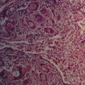
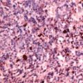
A biopsy specimen of the pigmented lesions indicated that WW has malignant melanoma.
