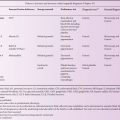
Table 3.1 Preliminary screening tests in blood or urine.
| Disease or group of diseases | Test | Technique |
| (a) Biochemical tests | ||
| MPS | Urinary glycosaminoglycans and derived oligosaccharides | Quantitative precipitation and qualitative electrophoresis or TLC; tandem mass spectrometry |
| Glycoproteinoses | Urinary oligosaccharides | TLC, GC-MS, tandem mass spectrometry |
| Sialic acid storage disease and sialidosis | Urinary free and total sialic acid | Colorimetric assay, tandem mass spectrometry |
| Glycosphingolipidoses | Urinary glycolipids | Tandem mass spectrometry |
| Pompe disease | Urinary oligosaccharides | Tandem mass spectrometry |
| Cobalmin-F transporter | Homocysteine and methylmalonic acid in blood and urine | Tandem mass spectrometry |
| (b) Histochemical tests | ||
| Gaucher | Gaucher cells, foamy macrophages and sea-blue histiocytes in bone marrow smears | Staining of bone marrow aspirate |
| Metachromatic leukodystrophy | Metachromatic granules (sulfatide) in urine | Stained urinary sediment |
| GM1-gangliosidosis (type 1), Galactosialidosis, sialic acid storage, sialidosis, fucosidosis, α-mannosidosis, I-cell disease, CLN3 | Numerous large and bold vacuoles in lymphocytes | Stained blood film |
| Niemann–Pick type A, Pompe, Wolman | Small discrete vacuoles in lymphocytes | Stained blood film |
| MPS VI & VII, multiple sulphatidosis | Alder granulation of neutrophils | Stained blood film |
| (c) Serum/plasma proteins | ||
| Gaucher disease, other lipidoses and neurodegenerative disorders | Chitotriosidase | Enzyme assay |
| Pompe disease | Creatine kinase | Enzyme assay |
| Mucolipidosis IV | Gastrin | Immunoassay |
Table 3.2 Enzymes that can be assayed initially in plasma*.
| Enzyme | Disease |
| Arylsulfatase A† | I-cell |
| Aspartylglucosaminidase | Aspartylglucosaminuria |
| α-Fucosidase† | Fucosidosis |
| α-N-acetylgalactosaminidase | Schindler |
| α-Galactosidase† | Fabry |
| β-Glucuronidase† | MPS VII |
| Total β-hexosaminidase† | Sandhoff |
| β-Hexosaminidase A† | Tay–Sachs / B1 variant |
| α- and β-Mannosidases | α- and β-Mannosidosis |
| Chitotriosidase§ | All children under 1 year and all with hepatosplenomegaly |
*Usually plasma but activities can be measured in serum
†Pseudodeficiency of enzyme reported
§Common null allele
Diagnosis of lysosomal enzyme defects
There is a deficiency of a specific lysosomal hydrolase in about 60% of the LSDs (see Classification in Chapter 5), which account for about 85–90% of the cases diagnosed. The diagnosis of a lysosomal enzyme defect is made by demonstrating a deficiency of a specific enzyme activity in white blood cells, lymphocytes or dried blood spots. Cultured fibroblasts are not used routinely as the primary source of enzyme activity unless the activity cannot be measured reliably in white blood cells, e.g. neuraminidase. Some enzyme deficiencies can be demonstrated reliably in plasma (Table 3.2). The enzymes are usually assayed in groups or panels (Table 3.3), according to the clinical indications (see Chapter 2) and the results of the preliminary tests. Some laboratories assay a large number of enzymes routinely, whereas others are very selective. Most lysosomal enzymes can be assayed using synthetic substrates, such as derivatives of the flurophore, 4-methylumbelliferone or the chromophore, paranitrophenol to increase the sensitivity and simplify the procedure. Novel substrates have been developed for use in mass spectrometric assays. They increase the sensitivity, permit simultaneous assay of several enzymes (multiplexing) and are particularly useful for measuring activity in dried blood spots [3, 5]. To validate a diagnosis another lysosomal enzyme must be assayed in the same sample at the same time to check the viability of the sample. Samples from a known case of the specific LSD (positive control) and from a normal control (negative control) should always be assayed at the same time under the same conditions. The activities are compared to reference ranges obtained under the same conditions. A complete deficiency or a very low amount of a specific enzyme activity is considered a definitive diagnosis of a specific LSD. The diagnosis is usually confirmed by DNA analysis. Some variant forms of LSD with a later onset and/or slower course have measurable residual enzyme activity with synthetic substrates. Their diagnosis can be confirmed by genetic testing because most variants are associated with specific mutations. Diagnostic laboratories should regularly participate in external quality assurance schemes [6, 7].
There are several complications with the diagnosis of enzyme defects that require special attention and experience.
Pseudodeficiencies
A polymorphism that lowers the enzymatic activity without causing disease can lead to a pseudodeficiency of a lysosomal enzyme activity and a false-positive diagnosis or ambiguous result. A pseudodeficiency has been found for at least nine lysosomal enzymes (listed in Classification in Chapter 5) but is particularly frequent for the arylsulfatase A enzyme, a deficiency of which causes metachromatic leukodystrophy (MLD) (see Chapter 9). Analysis of the patient’s DNA can distinguish between mutations causing MLD and the pseudodeficiency, which can also occur on the same chromosome as a disease-causing mutation. Discrepancies between the clinical indication and an apparent enzyme deficiency should alert the laboratory to the possibility of a pseudodeficiency.
Sphingolipid activator proteins defects
In contrast normal activity is sometimes found when attempting to diagnose a sphingolipidosis with synthetic substrates, despite strong clinical indications or manifestation of storage in preliminary tests. This could be due to a defect in a sphingolipid activator protein or SAP (see Chapters 6, 8 and 9). Five SAPs have been described: GM2 activator protein and saposins A–D derived from a common precursor, prosaposin. Whereas lysosomal hydrolases require a SAP to act on many glycolipids in vivo, they can act on soluble synthetic substrates in the absence of the saposin in vitro to give a false negative result if a SAP is defective. Variant forms of GM2-gangliosidosis, Gaucher disease, Krabbe disease and metachromatic leukodystrophy resulting from SAP deficiencies and a neurovisceral storage disease resulting from a deficiency of prosaposin have been described (see Classification in Chapter 5). A functional test, immunodetection of the SAP and/or a molecular genetic test are carried out to confirm the diagnosis.
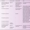
Table 3.3 Panels of enzymes assayed in blood according to clinical indication (see Chapter 2). The groups of enzymes in each panel may vary and are not prescriptive.
| A. Hydrops foetalis (see Figure 2.2) | |
| Disease | Enzyme Tests |
| MPS VII (very common) | β-Glucuronidase |
| Sialidosis (ML I) | α-Neuraminidase |
| Galactosialidosis | α-Neuraminidase/β-galactosidase |
| GM1-gangliosidosis | β-galactosidase |
| Gaucher | β-glucosidase |
| MPS I (Hurler) | α-Iduronidase |
| Wolman | Acid lipase |
| Niemann–Pick A | Sphingomyelinase |
| MPS IVA (Morquio A) | Galactosamine-6-sulfatase |
| Farber | Ceramidase |
| ML II (I-cell disease) | Multiple deficiencies in fibroblasts or increases in plasma |
| Other tests | |
| Infantile sialic acid storage | Sialic acid in amniotic fluid or urine |
| Niemann–Pick C | Cholesterol esterification |
| Plasma chitotriosidase is assayed in all cases | |
| B. Dysmorphic | |
| Disease | Enzyme |
| GM1-gangliosidosis | β-galactosidase |
| Multiple sulphatidosis | Arylsulfatase A |
| Fucosidosis | α-Fucosidase |
| α-Mannosidosis | |


