The classic triad for presentation of esophageal cancer is as follows:
■Asthenia
■Anorexia
■Analgesia (for dysphagia)
DIAGNOSIS
■Symptoms
•Dysphagia or odynophagia
•Hematemesis
•Dyspepsia
•Hoarseness
•Dyspnea
•Anorexia
■Signs (usually late presentation)
•Horner syndrome
•Left supraclavicular lymphadenopathy (Virchow’s node)
•Cachexia
•Hepatomegaly
•Bone metastases (rare, but paraneoplastic hypercalcemia can occur)
■Upper gastrointestinal endoscopy
•This diagnostic procedure is the gold standard. The combination of endoscopic biopsies and brush cytology has an accuracy of greater than 90% in making a tissue diagnosis of esophageal cancer.
■Barium contrast radiography
•This diagnostic procedure can document contour and motility abnormalities and unexpected airway fistula and may be useful when the entire esophagus has not been visualized endoscopically. However, a tissue diagnosis is needed for definitive diagnosis.
PATHOLOGY
■Most newly diagnosed patients have ADC, but there are contrasting reports on their relative prognosis. Less than 1% of esophageal tumors are lymphoma, melanoma, carcinosarcoma, or small cell carcinoma.
■Fifty percent of tumors arise in the lower one-third of the esophagus, 25% arise in the upper esophagus, and 25% of tumors occur in the middle one-third of the esophagus.
STAGING
The American Joint Commission for Cancer (AJCC) has designated staging of cancer by TNM classification, which defines the anatomic extent of disease. The most recent edition of this staging system proposes a distinction between T4a and T4b tumors—those that invade other structures but are resectable or unresectable, respectively. Other changes include staging based on histology and tumor grade, as well as N-staging based on the number of affected locoregional lymph nodes.
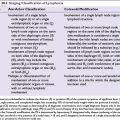
The Siewert classification subclassifies gastroesophageal junction tumors into three types according to their anatomic location: type I are distal esophagus tumors, type II are cardia tumors, and type III are subcardia gastric tumors.
Staging workup can include the following:
■Computerized tomography (CT) scan: CT scan of the chest and abdomen can demonstrate evidence of spread of tumors to lymph nodes or distant metastases to the liver (35%), lungs (20%), bone (9%), and adrenals (5%). CT scan may underestimate the depth of tumor invasion and peri-esophageal lymph node involvement in up to 50% of cases. Magnetic resonance imaging (MRI) provides similar results to CT.
■Endoscopic ultrasound (EUS): EUS may be helpful when metastases are not detected by CT or other imaging modalities. EUS is the optimal technique for locoregional staging. A meta-analysis demonstrated greater than 71% sensitivity in staging preoperative depth of invasion (T) and greater than 60% sensitivity for locoregional lymph nodes (N); specificity was greater than 67% and greater than 40%, respectively.
■Positron emission tomography (PET)/CT: PET/CT is useful when CT is negative for metastatic disease, and can change management of the disease in 25% to 40% of patients. It has limited utility in establishing T stage, and EUS is superior in establishing N stage, but PET/CT has a greater sensitivity for detecting distant metastatic disease than CT alone. PET/CT is currently being investigated for use in tailoring treatment based on metabolic response in the CALGB 80302 study.
■Bronchoscopy is required in tumors less than 25 cm from the incisors to exclude invasion of the posterior membranous trachea or tracheoesophageal fistula.
■Laparoscopy is sometimes performed for staging of EGJ tumors without evidence of metastatic disease, to rule out peritoneal dissemination.
TREATMENT
Surgery
■Surgery alone remains a recognized treatment for esophageal cancer with resectable local or locoregional disease. In 1993, surgery was used as a component of treatment in 34% of patients. Surgery alone was used in 18% of patients. In recent years, the improved survival seen with combined modality treatment has meant that surgery alone is generally only considered for patients with very early stage disease (T1-2N0M0).
■Endomucosal resection (EMR) with ablation is considered definitive treatment for patients with early T1a tumors, and may provide staging information for tumors with more advanced T stage.
■Recent improvements in staging techniques and patient selection have improved surgical morbidity and mortality. Operative mortality rates are now less than 5%. Surgical expertise is a major contributor to survival, with better outcomes in high-volume centers. Resection is possible in approximately 50% of patients. Five-year survival in patients with surgical resection is 5% to 25%.
■Surgical principles include a wide resection of the primary tumor with the goal of an R0 resection (no residual tumor), including more than 5 cm resection margins plus regional lymphadenectomy. Intraoperative frozen section can assess residual disease, which, if present, is considered an R1 (microscopic tumor) or R2 (macroscopic tumor) resection.
■In general, patients with cervical carcinoma of the esophagus (above the aortic arch) are not considered candidates for surgical resection; chemoradiation is favored in these patients. Other indicators of unresectable disease include T4 tumors or extensive nodal disease. Medical unresectability (due to comorbidities or poor performance status) is another common reason for patients not proceeding to esophagectomy.
■Surgical approaches include the following:
•Transthoracic resection: En bloc esophagectomy requires laparotomy and thoracotomy, for example, total thoracic or transthoracic (Lewis) procedures. A three-field lymph node dissection (extended lymphadenectomy) includes superior mediastinum and cervical lymphadenectomy. It is the treatment of choice in Japan, but is associated with increased toxicity and has a questionable survival advantage.
•Transhiatal esophagectomy: This includes laparotomy and cervical anastomosis. This technique avoids thoracotomy.
Chemoradiotherapy (Combined-Modality Approach)
Although no large prospective randomized trials have directly compared primary chemoradiation with surgery, definitive chemoradiation for locoregional carcinoma of the esophagus is considered an alternative to surgery.
Inoperable Disease
Definitive Chemoradiotherapy
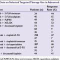
■The Radiation Therapy Oncology Group (RTOG) 85-01 trial demonstrated a survival advantage (14 vs. 9 months median survival and 27% vs. 0% 5-year survival) in favor of chemoradiotherapy over radiotherapy alone. The study used a regimen of cisplatin and short-course infusional 5-FU in combination with 50 Gy radiotherapy, compared with 64 Gy radiotherapy alone. A number of randomized trials of chemoradiotherapy versus radiotherapy alone have failed to duplicate the results of RTOG 85-01; however, a Cochrane review has confirmed the superiority of chemoradiotherapy versus radiotherapy in fit, motivated patients.
■The cisplatin/fluorouracil regimen used in the RTOG study carries significant toxicity, and alternative regimens have been sought to allow for effective treatment with better safety profiles. The recently presented phase III PRODIGE-5/ACCORD-17 study demonstrated similar survival in patients treated with a fluorouracil/oxaliplatin (FOLFOX) regimen to those treated with the RTOG regimen, but with lower rates of death from toxicity (1.1% vs. 6.4%) or within 15 days from chemotherapy (1.1% vs. 3.2%).
Operable Disease
Definitive Chemoradiotherapy Direct comparisons of chemoradiotherapy versus surgery in resectable esophageal cancer are limited and the optimal approach is controversial. Two trials have provided evidence to support a nonsurgical approach in some patients:
■In the first trial, patients with locally advanced but resectable squamous tumors were treated with chemoradiotherapy, and patients with at least a partial response were randomized to continued chemoradiotherapy or surgery. There was an improvement in local control rates (64% vs. 41% at 2 years), but no difference in overall survival, and early mortality and length of hospital stay were less in the chemoradiotherapy arm (12.8% vs. 3.5%, P = 0.03)
■The second trial randomized patients with locally advanced squamous tumors to either definitive chemoradiotherapy or chemoradiotherapy (lower doses of radiation) and surgery. There was no significant difference in survival outcomes (17.7 vs. 19.3 months, respectively) between the two groups of patients.
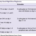
Radiation dose escalation has not proved to be beneficial. A trial examining this approach was closed after an interim analysis indicated that there would be no advantage with higher doses of radiation.
Neoadjuvant Chemoradiotherapy (Trimodality Approach)
■The rationale for preoperative chemoradiotherapy was first studied by Leichman et al. in 21 patients with SCC. The patients were treated with 30 Gy of radiation and with two cycles of concurrent 5-fluorouracil (5-FU) and cisplatin. An additional 20 Gy of radiation was given postoperatively when a residual tumor was seen at surgery. The pathologic complete response was 37%, with a median survival of 18 months.
■A number of prospective randomized phase 3 trials have addressed the issue of whether preoperative chemoradiotherapy offers any benefit over surgery alone. Much debate exists over interpretation of these trials.
Related posts:

Stay updated, free articles. Join our Telegram channel

Full access? Get Clinical Tree


