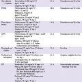
■From a family with a known deleterious BRCA1/2 mutation
■With a personal history of breast cancer and one of the following:
•Diagnosed below the age of 45
•Diagnosed below the age of 50 with 1 or more close blood relative with breast cancer younger than 50 and/or 1 or more blood relative with epithelial ovarian cancer at any age
•Two breast primaries when the first breast cancer diagnosis occurred at less than 50 years of age
•Diagnosed below the age of 60 with triple-negative breast cancer
•Diagnosed below the age of 50 with limited family history
•Diagnosed at any age with 2 or more close blood relatives (first-, second-, or third-degree relatives) with breast and/or epithelial ovarian cancer at any age
•Diagnosed at any age with two or more close blood relatives with pancreatic cancer at any age
•Close male blood relative with breast cancer
•Individuals of ethnicity associated with higher mutation frequency (i.e., Ashkenazi Jewish)
■With a personal history of epithelial ovarian cancer
■With a personal history of male breast cancer
■With a personal history of pancreatic cancer at any age with two or more close blood relatives with breast and/or ovarian and/or pancreatic cancer at any age
Testing of unaffected individuals should only be considered when no affected family member is available and family history reveals a first- or second-degree relative meeting the above criteria. In this circumstance, the significant limitations of interpreting results should be discussed since a negative test result in an unaffected individual can be uninformative. Testing consists of full sequencing of the BRCA1/2 genes as well as BART testing (BRCA analysis rearrangement test), which identifies large genomic rearrangements not picked up by routine sequencing.
If a BRCA mutation is found, the following recommendations should be implemented for women: breast self-examination training and education starting at age 18, clinical breast examination every 6 to 12 months starting at age 25, and annual mammography and MRI screening starting at age 25 or individualized based on earliest age of onset in the family. The addition of breast MRI to mammography increased the rate of breast cancer detection from 45% to 95% in one study, and most cancers were detected at stage 0 or 1. Prophylactic mastectomy can reduce the risk of breast cancer by 90% to 100%, and in those who decline surgery, chemoprevention with tamoxifen or raloxifene has been shown to reduce the risk of breast cancer by at least 50% and is a reasonable alternative along with close monitoring. Given the elevated risk for ovarian cancer and the lack of effective screening, risk-reducing salphingo-oopherectomy (RRSO) is recommended between the ages of 35 to 40, or upon completion of childbearing. Until then, a CA125 level and a transvaginal ultrasound should be considered every 6 months starting at age 30 or 5 to 10 years prior to the earliest age of diagnosis of ovarian cancer in the family.
For men, the risk of breast cancer is 2% to 8% and is usually seen after age 50. Breast self-examinations should start at the age of 35 and baseline mammography can be considered at the age of 40 and continued only if gynecomastia or parenchymal/glandular breast density is present. Chemoprevention and prophylactic mastectomies are not offered to men because the incidence of breast cancer is not nearly as high as in women. Male carriers also have a higher risk for both prostate and pancreatic cancer than the general population. Prostate cancer screening should start at age 40 with a baseline digital rectal examination (DRE) and PSA level and repeated per the NCCN guidelines which can be reviewed separately.
For both men and women, annual skin examinations are recommended given the increased risk of melanoma, and the general population screening guidelines for colon cancer prevention should be followed. For pancreatic cancer prevention, alcohol and tobacco use should be avoided. Screening can be offered to individuals with a strong family history with either EUS or MRI every 1 to 3 years and an annual CA 19-9 level. Studies looking at pancreatic cancer screening are limited and the utility of screening is not completely known.
Cowden Syndrome
Cowden syndrome is an autosomal dominant syndrome with an incidence of 1 in 200,000. It is caused by a loss of function in the tumor suppressor PTEN gene and is associated with multiple hamartomas in a variety of tissues, characteristic dermatologic manifestations, and an increased risk of breast, endometrial, thyroid, kidney, melanoma, and colorectal cancers. The lifetime risk of breast cancer is 25% to 50%, although it may be as high as 85%. Thyroid cancer develops in two-third of carriers and can occur in childhood. Pathology is usually follicular, rarely papillary, and never medullary. Renal cell carcinoma (RCC) can be seen in 13% to 34% of carriers. The prevalence of colon polyps is 66% to 93% and they are usually hamartomatous or inflammatory polyps with a lifetime risk of 16% for colon cancer. Neurologic manifestations include dysplastic gangliocytoma of the cerebellar cortex, macrocephaly, and mental retardation/developmental delay/autism. Women commonly have benign abnormalities such as breast hamartomas, uterine fibroids, and ovarian cysts. Men often have lipomatosis of the testes seen as hyperechoic lesions on testicular ultrasound. Both men and women frequently have benign thyroid lesions, such as adenomas and multinodular goiter. Benign esophageal acanthosis can also be seen.
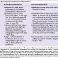
The diagnostic criteria are divided into pathognomic criteria, major criteria, and minor criteria. The pathognomic criteria include mucocutaneous lesions such as facial trichilemmomas (skin tags), acral keratoses (thickened area of skin that may be red, yellow, or brown), or pappilomatous oral lesions (small wart-like growths). The major criteria include breast cancer, thyroid cancer, macrocephaly, endometrial cancer, and dysplastic gangliocytoma of the cerebellar cortex. The minor criteria include benign structural thyroid disease, mental retardation, hamartomatous gastrointestinal (GI) tract polyps, benign cystic breast disease, lipomas or fibromas, and genitourinary tract tumors. Both the NCCN and the International Consortium Cowden Consortium have criteria for testing high-risk individuals and guidelines for the care of carriers. In the absence of family history, Cowden syndrome is diagnosed if any of the following are present: six or more facial papules including at least three trichilemmomas, facial papules and oral pappilomatous, oral pappilomatous and acral keratoses, or six of more acral keratoses on the hands and feet. One major and three minor or four minor criteria are also considered diagnostic. If there is a family history of Cowden syndrome, then one pathognomonic, one major, or two minor criteria are diagnostic. Only 20% to 34% of individuals who meet these criteria have germline PTEN mutations. PTEN testing includes sequencing of the entire coding region and deletion/duplication analysis. Mutations have also been reported in the PTEN promoter region and in other genes including succinate dehydrogenase (SDH) subunits B and D.
The management guidelines for women with Cowden syndrome include breast self-examination training and education starting at age 18, clinical breast examination every 6 to 12 months starting at age 25, and annual mammography and MRI screening starting at age 25 or individualized based on earliest age of onset in family. There are no specific guidelines for endometrial cancer screening and carriers should be educated and followed closely, with a prompt response to symptoms. Risk-reducing mastectomies and hysterectomy can be considered. Men and women should have an annual physical examination starting at age 18 or 5 years prior to the youngest age of diagnosis of cancer in their family with emphasis on the breast and thyroid examination. Baseline thyroid ultrasound should be done at age 18 and annually after that. Screening colonoscopies starting at age 35, annual dermatologic examinations, and education about the signs and symptoms of cancer can be considered.
Li–Fraumeni Syndrome
Li–Fraumeni syndrome (LFS) is a hereditary syndrome associated with a wide range of cancers that appear at an unusually young age. It has an autosomal dominant pattern of inheritance and is associated with mutations in the p53 tumor suppressor gene, which plays a major role in DNA repair. The absence of this gene allows for the survival and proliferation of cells with damaged DNA. The lifetime risk of cancer is nearly 100%, with 90% of individuals diagnosed with cancer by age 60. The classic tumors seen in this syndrome are sarcoma, breast cancer, leukemia, brain tumors, and adrenal gland cancers.
Classic Li–Fraumeni criteria include a proband with sarcoma before the age of 45, a first-degree relative with cancer before the age of 45, and a first- or second-degree relative with cancer before the age of 45 or sarcoma at any age. Chompret criteria include one of the following:
■A proband who has a tumor belonging to the LFS spectrum (sarcoma, premenopausal breast cancer, brain tumor, adrenocorticoid tumor, leukemia, or lung bronchoalveloar cancer) before age 46 and at least one first- or second-degree relative with a tumor in the LFS spectrum before age 56 or with multiple tumors.
■A proband with multiple tumors (except multiple breast tumors), two of which belong to the LFS spectrum and the first of which occurred before age 46.
■A proband who is diagnosed with adrenocortical tumor or choroid plexus tumor regardless of age irrespective of family history.
Testing of individuals who meet either of these criteria or women with breast cancer before age 35 who have tested negative for the BRCA1/2 mutations is recommended.
The management guidelines for women with LFS include breast self-examination training and education starting at age 18, clinical breast examination every 6 to 12 months starting at age 25, and annual mammography and MRI screening starting at age 25 or individualized based on earliest age of onset in family. All carriers should have an annual physical examination including skin and neurologic examinations. Radiation therapy should be avoided if possible (i.e., mastectomy instead of lumpectomy for breast cancer) due to the increased risk of radiation-induced malignancies. Colonoscopy screening should start no later than age 25. Other options for screening should be discussed with the patient such as whole-body MRI, abdominal ultrasound, and brain MRI. Targeted surveillance should be based on family history.
HEREDITARY GI SYNDROMES
Lynch Syndrome
Lynch syndrome, also known as hereditary nonpolyposis colorectal cancer (HNPCC), is an autosomal dominant disorder characterized by germline mutations in DNA mismatch repair (MMR) genes. The absence of these genes results in a significantly increased risk of cancer formation of up to 80%, with the most common carcinomas located in the colon, rectum, and uterus.
Lynch syndrome accounts for 2% to 3% of all colon cancers with a lifetime risk of up to 70%. Compared to those with sporadic colon cancer, HNPCC patients are usually younger in age (45 vs. 65 years old) and have more poorly differentiated, mucinous tumors found in the right colon. Despite these more aggressive histologic features, affected patients have better 5-year survival rates compared to those with common sporadic colorectal cancer, likely due to the lower risk of metastases. Though uncommon, a small subset (10%) of HNPCC patients will have synchronous (two primary tumors) or metachronous (second tumor developing at least 6 months after the first) cancers.
Endometrial carcinoma is the most common extracolonic tumor in Lynch syndrome, accounting for about 2% of all uterine cancer and with a reported incidence as high as 70% in female carriers. Similar to colon cancer in Lynch syndrome, women are typically younger in age at diagnosis (50 vs. 60 years old). Other sites at increased risk of cancer development include the ovaries, stomach, small bowel, pancreas, hepatobiliary system, upper urinary tract (renal pelvis and ureter), skin, and brain. The chances of developing cancer in these various organs vary depending on which gene is mutated, but the incidence can be as high as 20%.
Related posts:

Stay updated, free articles. Join our Telegram channel

Full access? Get Clinical Tree


