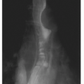
Saucerization of Pressure Damage
At pressures above the capillary filling pressure of 32 mm Hg, arteriolar perfusion is jeopardized. Pressure at the skin surface has a cone-shaped radiation, resulting in much greater internal distribution. The skin is actually less susceptible to damage than the subcutaneous tissue is. Subcutaneous tissue, in turn, is better protected than muscle, which is most vulnerable to pressure damage. Therefore, on examining the skin we frequently see just the tip of the iceberg as far as tissue damage.
11The multidisciplinary National Pressure Ulcer Advisory Panel (NPUAP), formed in 1987, is an independent, nonprofit organization established to evaluate areas of concern in pressure ulcer management. NPUAP’s pressure ulcer staging system,
12 the most frequently used, has been adopted by the AHRQ guideline panels. The NPUAP staging system is:
Stage I:
Pressure ulcers are observable pressure-related alterations of intact skin whose indicators, as compared to adjacent or opposite body areas, may include changes in skin temperature (i.e., warmth or coolness), tissue consistency (i.e., firmness or bogginess), and/or sensation (i.e., pain, itching). The pressure ulcer appears as a defined area of persistent redness in lightly pigmented skin, whereas in darker skin tones, the pressure ulcer may have persistent red, blue, or purple hues.
Stage II:
Pressure ulcers are associated with partial-thickness skin loss involving epidermis, dermis, or both. The ulcer is superficial and presents clinically as an abrasion, blister, or shallow crater.
Stage III:
Pressure ulcers are associated with full-thickness skin loss involving damage to, or necrosis of, subcutaneous tissue that may extend down to, but not through, the underlying fascia. The pressure ulcer presents clinically as a deep crater, with or without undermining of the adjacent tissue.
Stage IV:
Pressure ulcers are associated with full-thickness skin loss with extensive destruction, tissue necrosis, or damage to muscle, bone, or supporting structures (e.g., tendon, joint, or capsule). Undermining and sinus tracts may also be associated with Stage IV pressure ulcers. (The above is adapted from National Pressure Ulcer Advisory Panel (NPUAP). NPUAP position on reverse staging of pressure ulcers, in Advanced Wound Care, Vol. 8. 1998:32).
Because skin is a not a uniform organ, its appearance or thickness varies according to anatomic site. On average, a pressure ulcer that is approximately 2 mm or deeper and is
located on the trunk, pelvic girdle, ankle, or heel is usually classified at least as a Stage III. This depth approximates that of a US nickel or a house key. Initial pressure ulcer staging is important because the ulcer is not restaged as it heals.
Reverse (Healing) Staging
The NPUAP has issued a position statement
12 against using back staging or reverse staging. For example, a healing Stage IV pressure ulcer would not be progressively downstaged to III, II, and so on, as it heals. A healing pressure ulcer is replaced with scar tissue because dermis, subcutaneous fat, and muscle cannot be regenerated. Therefore, restaging as an ulcer heals does not describe the physiologic changes that occur. If a previously healed pressure ulcer reopens, the prior staging diagnosis should be used (i.e., once an ulcer is Stage IV, it will always remain Stage IV).
NPUAP recommends
12 that pressure ulcers also be described by size, the presence of granulation tissue, and the presence of infection and that an appropriate pressure ulcer healing tool (e.g., the Pressure Ulcer Scale for Healing [PUSH] tool [see
Fig. 37.2]) also be used.


