Pros
Cons
No radiation
Claustrophobia
Multiplanar
Non-MRI-compatible metal
Excellent soft tissue details
Cost
Better resolution
Tissue differentiation
MRI Protocol
T1 sagittal
Dual-echo axial
Pre- and post-contrast high-resolution T1 sagittal and coronal to pituitary gland (1 mm or less)
T2 WI coronal to pituitary gland
Diffusion-weighted scan – optional
MR angiogram – optional
CT Scanning
Pros | Cons |
|---|---|
Availability | Radiation exposure (reduced with cone beam CT scanners) |
Fast | Soft tissue detail inferior to MRI |
Relatively inexpensive | |
Good bone detail | |
Acute bleed/proteinaceous material | |
Surgical planning – navigation |
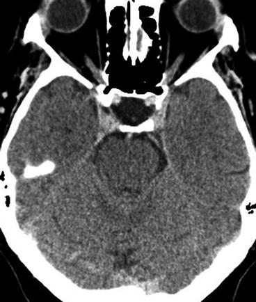
CT scans can provide additional information about the bony margins of the fossa. It may be useful in identifying bone asymmetry, expansion, or erosion if present. Calcification is also easier to identify on CT (Fig. 38.1).