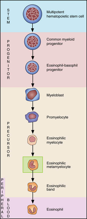
7 Eosinophil maturation
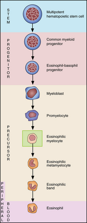
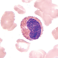
Eosinophilic myelocyte
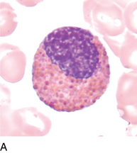
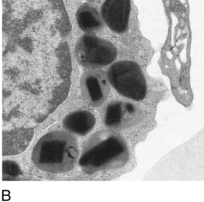
FIGURE 7–2B Electron micrograph of eosinophilic granules to demonstrate crystalline structure of granules.
7 Eosinophil maturation
FIGURE 7–2B Electron micrograph of eosinophilic granules to demonstrate crystalline structure of granules.