Qualitative Disorders of Platelet Function


Qualitative Disorders of Platelet Function


Thomas J. Kunicki

Diane J. Nugent


Much of our current understanding of normal platelet structure-function relationships has been derived from the study of patients with congenital platelet disorders. These syndromes are the subject of excellent recent reviews.1, 2, 3, 4, 5


DIAGNOSIS AND CLASSIFICATION OF PLATELET DYSFUNCTION: AN ALGORITHM

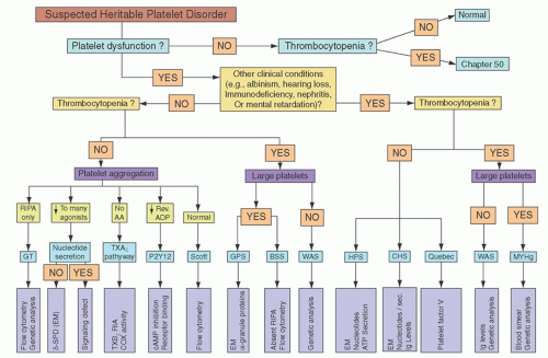
The preliminary diagnosis of platelet dysfunction must be made on the basis of patient history, and the diagnosis then confirmed by specific laboratory tests of platelet function. A practical algorithm for this purpose, depicted in Figure 52.5, is reproduced with permission from the excellent review by Bolton-Maggs et al.5


Bedside Exam and Patient History

An accurate and detailed patient and family history is a key element in the assessment of platelet disorders. One should bear in mind that bleeding histories are subjective and that bleeding can be variable, often evolving or decreasing throughout a person’s lifetime. In particular, children may not have yet had enough hemostatic challenges to develop a strong clinical. The subjectivity of the oral history is reflected in a recent statistic that at least one quarter of persons who complain of serious bleeding do not have a bleeding disorder, whereas at least one third of persons who
have no bleeding can be shown to have von Willebrand disease (vWD) or a platelet disorder.6


FIGURE 52.2. Initiation of platelet adhesion by matrix components, particularly collagen. Platelets employ a number of collagen receptors. These include VWF-mediated binding of collagen to the glycoprotein Ib (GPIb) complex (a heptamer composed of GPIbα, GPIbβ, GPV, and GPIX), the direct engagement of collagen by the integrin α2β1 and GPVI/FcRγ. Engagement and clustering of GPVI initiates tyrosine phosphorylation of FcRγ by a Src family kinase (SFK). The tyrosine kinase Syk then binds and is activated, in turn activating phospholipase Cγ, which then initiates phosphoinositide hydrolysis, secretion of ADP, and the production of TXA2. ADP and TXA2 augment platelet activation by binding to their respective platelet receptors.


FIGURE 52.3. The extension phase of platelet plug formation accelerates and augments the activation of the platelet and is mediated largely by G protein-coupled receptors, including: the purinogenic receptors P2Y1 and P2Y12, which are bound by ADP; the α and β isoforms of the thromboxane A2 (TXA2) receptor TP; the protease-activated receptor (PAR) family members PAR1 and PAR4, that are recognized by thrombin; and the α2A-adrenegic receptor that is specific for epinephrine.

To overcome the subjectivity of a history for superficial mucocutaneous bleeding, the International Society on Thrombosis and Hemostasis (ISTH) suggests that bleeding should be considered clinically significant when there are two or more distinct bleeding sites such as the skin, nose, gums, vagina, gastrointestinal tract, or genitourinary tract. This includes either spontaneous bleeding or provoked bleeding, such as that which might result from dental work, parturition, trauma, or surgery. In addition, a bleeding history involving only a single site should be considered significant when it is so severe as to warrant blood transfusions. Finally, a single bleeding symptom that recurs on three or more unrelated and separate occasions should also be considered significant.7, 8


FIGURE 52.4. The consolidation phase of platelet thrombus formation occurs through the bridging of adjacent integrin αIIbβ3 complexes by fibrinogen or VWF, as well as other adhesive proteins.


TABLE 52.1 DISORDERS OF PLATELET FUNCTION


Hereditary platelet dysfunction


Initiation phase


Bernard-Soulier syndrome


GPVI deficiency


Extension phase


Secretion disorders/granule deficiencies


α-Granule abnormalities (gray platelet syndrome)


δ-Granule (dense body) abnormalities


Hermansky-Pudlak syndrome


Chediak-Higashi syndrome


Wiskott-Aldrich syndrome


α/δ-Granule deficiency


Defects of signal transduction and secretion


Impaired liberation of arachidonic acid


Cyclooxygenase deficiency


Thromboxane synthetase deficiency


Thromboxane A2 receptor abnormalities


Defects in calcium mobilization


Defects of platelet procoagulant activity


Consolidation phase


Glanzmann thrombasthenia


Miscellaneous


Hereditary macrothrombopathy/sensorineural hearing loss


Acquired disorders of platelet function


Drug-induced platelet dysfunction


Analgesics


Antibiotics


Cardiovascular drugs


Psychotropic drugs


Secondary platelet dysfunction


Uremia


Paraproteinemia


Myeloproliferative disorders


A number of quantitative approaches to assess the relative severity of patient bleeding have been proposed, some including a normalization based on patient age.9, 10 However, such approaches work best for a comparison of cohorts of related individuals with comparable bleeding disorders, such as families of patients with vWD.

The distinction between normal individuals and those with bleeding disorders is not always clear cut. Care should be taken in all patients to note the use of medicines, either “over the counter,” herbal, or prescription, that are known to influence platelet function. This is particularly true of medications such as aspirin, other nonsteroidal anti-inflammatory drugs (NSAIDs), ticlopidine or clopidogrel, integrin αIIbβ3 antagonists (e.g., Abciximab, tirofiban, and eptifibatide), epoprostenol, statins, cilostazol, sildenafil, fluoxetine, and large doses of various β-lactam antibiotics (penicillins > cephalosporins).7

Bleeding manifestations typical of platelet dysfunction include: (1) Unexplained or extensive bruising; (2) epistaxis, particularly if lasting more than 30 minutes, causing anemia or admission to hospital; (3) menorrhagia, particularly if this has been present since the menarche; (4) oral cavity bleeding; (5) bleeding during
childbirth; (6) bleeding following invasive procedures; and (7) bleeding following dental extraction.


FIGURE 52.5. A scheme for the analysis of patients with suspected hereditary platelet dysfunction. In order to establish a precise diagnosis, specific laboratory tests are needed to establish a defect in platelet number or function. Generally, an automated full blood count, blood film, platelet aggregometry, and quantification of platelet nucleotides are necessary. The purpose of this algorithm is to assist the investigator in the systematic interpretation of laboratory results. To complete a diagnosis, specialty analyses may be necessary, and these are included for the specific disorder. Such assays might include flow cytometry to measure expression of surface glycoproteins, such as αIIb or β3 (GT), GPIb or GPIX (BSS). In addition, quantitation of annexin V binding (Scott syndrome) and genetic analyses to identify specific gene mutations might be needed: αIIb or β3 (GT), WAS (WAS and XLA) and MYH9. Certain disorders can be caused by mutations in more than one gene (e.g., HPS and BSS), so genetic analysis has not been considered as a definitive test in all cases. The algorithm includes the most frequent or best characterized heritable platelet disorders. Symbols and abbreviations: ↓, reduced aggregation; AA, arachidonic acid; BSS, Bernard-Soulier Syndrome; CHS, Chediak-Higashi syndrome; COX, cyclooxygenase; δ-SPD, dense-granule disorder; EM, electron microscopy; GPS, gray platelet syndrome; GT, Glanzmann thrombasthenia; HPS, Hermansky-Pudlak syndrome; MYH9, MYH-9-related disorder; P2Y12, deficiency of P2Y12 ADP receptor; QBS, Quebec platelet syndrome; Rev., reversible aggregation; RIA, radioimmunoassay; RIPA, ristocetin-induced platelet aggregation; Sec., secretion; TXA2, thromboxane A2; WAS, Wiskott-Aldrich syndrome. From Bolton-Maggs PH, Chalmers EA, Collins PW, et al. A review of inherited platelet disorders with guidelines for their management on behalf of the UKHCDO. Br J Haematol 2006:603-633.

Severe platelet dysfunction is present from early childhood onward. Following delivery of an affected infant one may find intracranial and/or subdural hemorrhage, excessive bleeding from the umbilical stump or after circumcision, or easy bruising after handling. As the infant becomes more mobile, easy or extensive bruising following relatively mild trauma can be indicative of platelet dysfunction. Prolonged epistaxis is a common finding throughout childhood and can even become life threatening. In adults, menorrhagia and bleeding during childbirth are common and potentially serious, whereas bleeding following any invasive procedure should be anticipated.5

Mild platelet dysfunction is more likely to manifest itself at any age most commonly following a definable hemostatic challenge, such as surgery or dental extractions. Easy bruising is a very nonspecific symptom, and many cases are difficult to distinguish from what would otherwise be considered a normal response.

Consanguinity increases the likelihood of an autosomal recessive platelet disorder, and a family history is invaluable in establishing the diagnosis of inherited platelet dysfunction.


Laboratory Assessment

A number of laboratory-based evaluations are critical for an accurate diagnosis of platelet dysfunction.


Whole Blood Platelet Count

A key element in assessing platelet dysfunction is an accurate whole blood platelet count. Automated counts should be viewed as provisional. Inasmuch as macrothrombocytes or platelet aggregates created inadvertently during blood processing will not be counted, the whole blood platelet count should be confirmed by an optical method.


Global Coagulation Tests

To rule out abnormalities in clotting factors, all patients should have a prothrombin time, activated partial thromboplastin time, and thrombin time performed. Laboratories should determine their own age-related normal range. It is also critical to investigate all patients for VWD, which is far more common than platelet function disorders and creates a similar bleeding phenotype.


Bleeding Time

The bleeding time is historically a common measure of platelet function. However, because the test is poorly reproducible, time consuming, and insensitive, the bleeding time has gradually fallen out of favor as a clinically useful test.11 In cases of mild platelet dysfunction, the bleeding time is often normal or minimally prolonged; 11 in severe cases, it will usually be prolonged.
Unfortunately, the bleeding time does not correlate well with the in vivo bleeding tendency within individual patients, and an accurate bleeding history is considered by many to be a more valuable screening test. Nonetheless, a prolonged bleeding time should be considered sufficient grounds to perform additional tests of platelet function.


Platelet Function Analyzer-100

The platelet function analyzer-100 (PFA-100) measures the rate of thrombus formation under high shear in citrated whole blood that is perfused through a membrane aperture coated with collagen/epinephrine or collagen/ADP. The closure time (CT) will be significantly prolonged in Glanzmann thrombasthenia (GT) and Bernard-Soulier syndrome (BSS) using either ADP/collagen or epinephrine/collagen membrane cartridges.12, 13 Consequently, the PFA-100 can be used to screen patients to exclude these diagnoses.

The PFA-100 may be sensitive to platelet storage pool disease (SPD), primary secretion defects, the Hermansky-Pudlak syndrome (HPS), and the Quebec syndrome. However, because falsenegative results occur in patients with all of these disorders, there are those who question its usefulness as a screening tool.12, 14 The PFA-100 is affected by platelet count and hematocrit, and is dependent on normal VWF levels and naturally occurring (genetic) differences in platelet membrane GPVI or α2β1 expression.13, 14, 15


Platelet Aggregation

Platelet aggregation in platelet-rich plasma remains an important test in the analysis of platelet function. The typical agonists that are used to induce platelet aggregation are ADP, epinephrine, collagen, arachidonic acid (AA), ristocetin, the TX receptor agonist U46619, thrombin or the thrombin receptor-activating peptide. Because the level and/or activity of each of the receptors for the agonists can vary among normal subjects, it is recommended that dose-response curves to each agonist be obtained from the patient under study and compared to a reference range obtained from multiple normal subjects.16 When thrombin is the agonist, an inhibitor of fibrin polymerization, such as glycine-proline-arginine-proline peptide, must be added to the PRP, or alternatively, plasma-depleted washed platelets must be used.

Platelet aggregation is sensitive to platelet count, and at counts ≤120,000/µl, the response to some agonists will be impaired. In thrombocytopenic samples, the best options are to adjust a control sample to the same count as the patient or to perform studies on washed platelets where the platelet number can be normalized. Consideration should also be given to more specialized tests, such as a measure of P-selectin expression or integrin αIIbβ3 activation by flow cytometry. The expected aggregation responses associated with specific diagnoses are covered in the appropriate sections.


Adenine Nucleotide Content and Release

Measurement of platelet adenosine nucleotide (ADP and adenosine triphosphate [ATP]) content and release can be used in the diagnosis of storage pool and release defects.17 A finding of normal platelet aggregation does not exclude the diagnosis of SPD. It is recommended that patients suspected of having platelet dysfunction should have both platelet aggregation and adenine nucleotide release performed, unless it is certain that the laboratory performing the platelet aggregation assays can demonstrate that their assay conditions are sensitive to defects in platelet nucleotide amount or release.

Platelet nucleotide content and release varies with age. Ideally, age-related normal ranges should be established for total and released levels of ATP and ADP and their ratios. Platelet aggregation, nucleotide content, and nucleotide release in children over 12 months of age do not differ significantly from adult values, whereas collagen-induced platelet nucleotide release has been shown to be reduced in neonates compared with children older than 1 year. Agonist-induced secretion of platelet granule contents has been shown to be reduced in both term and premature babies due to immature signal transduction pathways.18, 19


Flow Cytometry

Flow cytometry is routinely used to measure platelet surface receptor density, platelet activation, α-granule release, procoagulant phospholipid expression, and microvesicle production.20, 21 A common application of flow cytometry is the assessment of GPIb complex and integrin αIIbβ3 expression in the diagnosis of BSS and GT. Individuals who are heterozygous for these disorders are also readily distinguished. An important benefit of flow cytometry is the small quantities of blood required, an attractive feature in young children or thrombocytopenic individuals.


Electron Microscopy

Transmission electron microscopy (TEM) of fixed/embedded platelet thin sections can be performed by a limited number of specialized personnel, but is critical in the assessment of platelet granule defects and changes in platelet ultrastructure (e.g., in the evaluation of patients with MYH-9 defects). Whole-mount EM can be used to quantitate δ-granule content because this is readily identified in unstained preparations.22


Additional Assays

Several additional assays are available in specialized laboratories that can provide further information relevant to the diagnosis of the particular platelet disorder, including analysis of receptor expression, specific molecular or genetic defects, protein phosphorylation, formation of signal transduction intermediates, or a characterization of the platelet proteome. The utility of these tests in clinical diagnoses is currently under intense investigation.


Overview of Treatment Options for Platelet Disorders

Although the number of clearly identifiable platelet dysfunction syndromes is growing, our choice of treatment options remains limited. Minor membrane bleeding may be controlled with topical agents in the nasal or oral cavities with antifibrinolytic agents. Epsilon-aminocaproic or tranexamic acids will decrease blood loss associated with epistaxis or menorrhagia. Some patients may benefit from Stimate as documented by DiMichele and Hathaway,23 whereas others actually bleed more with this agent due to the fibrinolysis induced by this medication. For this reason many centers recommend the use of Stimate in conjunction with Amicar or Cyklokapron.

In the case of menorrhagia, hormonal suppression is the mainstay for women who do not wish to undergo endometrial ablation for hysterectomies. In combination with antifibrinolytics and Stimate, even severe bleeding may be controlled; however, more aggressive therapy including platelet infusion may be required to control bleeding before these agents take effect.

Platelet transfusion may pose a dilemma to those physicians wishing to avoid alloimmunization in patients who may require multiple transfusions throughout their lifetimes to control hemorrhage. This is particularly true for those patients who are lacking membrane GPs, such as αIIbβ3 or GPIb. In this setting patients are at risk of developing isoimmunization, making antibodies against the “foreign” proteins that they lack and thus becoming refractory to all subsequent platelet transfusions.


Although rare, this creates a significant challenge in patients who require frequent platelet infusions to control life-threatening bleeding. Patients with GT have not only developed antibodies to αIIb and/or β3 but also demonstrated anti-idiotypic antibodies that bind to fibrinogen, thus creating a hemorrhagic disorder far worse than the underlying platelet dysfunction. In general, physicians avoid platelet transfusion apart from cases of severe hemorrhage. Isoimmunization appears to be rare in Glanzmann but the risk of alloimmunization is still a major concern, therefore leukocyte depletion of transfused platelets is recommended to decrease the frequency of sensitization.

Activated recombinant Factor VII (rFVIIa) has been used to slow or arrest bleeding associated with platelet dysfunction.24 Dosages have varied widely but many patients have responded to this regimen when others have failed. Used in combination with antifibrinolytics, minor bleeding can be controlled in certain patients. This treatment is often used prior to platelet transfusion in order to avoid blood product exposure and isoimmunization.

For those patients who present with recurrent life-threatening bleeds, bone marrow transplant or stem cell infusion following immune ablation is recommended before the patients have extensive blood product exposure.25 Successful transplantation with normal stem cells represents long-term cure for these patients. Although complications related to stem cell transplantation cannot be overlooked, successful engraftment essentially eliminates the significant risk of mortality related to hemorrhage in patients with severe disorders.

Patients with rare platelet dysfunction syndromes are now included in many of the rare bleeding disorder international and regional registries that will aid physicians in understanding the natural course, optimal therapy, and life expectancy for each of the syndromes listed below, as well as the numerous platelet disorders that have yet to be defined. Definitive molecular and biochemical diagnoses will dictate appropriate therapy in these patients. With improvement in platelet function measurement and proteomic approaches one should see significant improvement in early diagnosis and medical management.


HEREDITARY DISORDERS OF PLATELET FUNCTION


Defects in the Initiation Phase: Bernard-Soulier Syndrome

BSS is a rare disorder first described in 1948 as “dystrophie thrombocytaire hemorrhagipare congenitale” caused by abnormal expression or activity of the platelet GPIb complex.26


Etiology

BSS platelets have a quantitative or qualitative abnormality of the membrane GPIb complex, a heptamer composed of four leucine-rich GP that are the products of distinct genes (Fig. 52.2). The prominent member of the complex, GPIb, is a heterodimer composed of disulfide-bonded GPIbα and GPIbβ subunits. GPIb then forms a noncovalent complex with GPIX, and two GPIb-IX trimers then associate noncovalently with one molecule of GPV. The amino-terminal type A domain of GPIbα binds directly to VWF, mediating normal platelet adhesion during the initial phases of primary hemostasis, whereas BSS platelets do not adhere to the extracellular matrix when perfused at a high shear rate.27 The defect in the GPIb complex also explains the failure of affected platelets to agglutinate in the presence of ristocetin, even in the presence of normal plasma or VWF.

Mutations in the gene for GPIbα, GPIbβ, or GPIX, but not GPV, have been shown to result in decreased expression of the GPIb complex and the BSS. Defects range from virtually absent GPIb to variant forms in which patients retain measurable amounts of apparently dysfunctional GPs.1

BSS is usually inherited as an autosomal-recessive disorder, and consanguinity is common in reported kindreds. Heterozygotes typically have “giant” platelets and reduced levels of the GPIb complex and may or may not be symptomatic.


Clinical Features

Bleeding symptoms are usually evident shortly after birth or in early childhood. The clinical manifestations include purpura, epistaxis, gingival bleeding and menorrhagia, and more rarely gastrointestinal bleeding, major hematomas, or hematuria. Severe bleeding episodes can result from trauma and surgical procedures, such as tonsillectomy, appendectomy, splenectomy, oral surgery, and menses. However, individual bleeding can vary substantially in severity and frequency.


Laboratory Findings

The typical laboratory findings include an increased bleeding time, mild thrombocytopenia, giant platelets on blood smear, and defective adhesion to collagens in vitro. Platelet morphologic abnormalities are a hallmark of BSS, featured by large platelets with a diameter as high as 10 µm. A defective platelet agglutination in response to ristocetin, as measured by aggregometry in vitro, is a unique characteristic of BSS that differentiates this syndrome from other rare inherited disorders that are also associated with macrothrombocytopenia, such as the MHY9-related disorders.1 A firm diagnosis requires the combined findings of increased bleeding times, macrothrombocytopenia, defective ristocetin-induced agglutination, and low or absent levels of platelet GPIb-V-IX (CD42a-d) by flow cytometry.

In bone marrow aspirates, megakaryocytes are normal or increased in number, but they reveal no characteristic morphologic abnormalities when viewed by light microscopy. Electron microscopic studies have revealed abnormalities of the dense tubular system and vacuolization of the demarcation membrane system.28


Management

Therapeutic approaches include both general and specific treatment of bleeding. Patients should be warned to avoid trauma and antiplatelet medication, such as aspirin, and to maintain proper dental hygiene. Females may benefit from contraceptive therapy once they reach puberty. Treatment of bleeding or prophylaxis during surgical procedures usually requires blood or platelet transfusion with the associated risk of developing antiplatelet alloantibodies. Desmopressin and rFVIIa administration have been shown to shorten the bleeding time in some patients. In rare cases of life-threatening bleeding, a bone marrow or umbilical cord hematopoietic stem cell transplantation may be considered.29 Responses to antifibrinolytic agents are more variable, and the administration of adrenal corticosteroids and splenectomy are usually ineffective.

Platelet-reactive isoantibodies have been generated by BSS who have received multiple blood or platelet transfusions.30 These antibodies produce a particularly severe clinical complication because they will bind to and may neutralize the function of GPIb on transfused platelets. This results in impaired adhesion of the transfused platelets. The presence of such antibodies can be established by a finding of impaired in vitro aggregation of normal platelets induced by ristocetin and bovine VWF in the presence of patient plasma. Alloimmunization is also a common side effect of multiple platelet transfusions, and this may necessitate the subsequent use of HLA-matched platelets.


Variation in the Initiation Phase: Collagen Receptor Polymorphisms

A lifelong bleeding disorder and the unique absence of in vitro platelet aggregation to collagen have been associated with a deficiency of either of the collagen receptors, integrin α2β1 or GPVI,1 yet specific gene defects have not been identified. Careful evaluation is needed to establish a specific molecular defect in either receptor.


The Integrin α2β1

Among normal individuals, platelet α2β1 levels can vary up to tenfold and correlate with differences in adhesiveness to type-I or type-III collagens and genetic variants of the α2 subunit gene ITGA2. We identified several single nucleotide polymorphisms (SNPs) within the coding sequence of the α2 gene ITGA2 that correlate with platelet α2β1 density31 and define six major ITGA2 haplotypes.


Clinical Relevance of α2 Polymorphisms

In VWD, platelet adhesive functions are impaired due to the decrease in functional VWF multimers in plasma and platelets. Inheritance of the ITGA2 haplotypes associated with lower α2β1 density will increase the risk of bleeding in patients with either type I and type2 VWD.9, 10

The association of ITGA2 haplotypes with risk for arterial thrombosis has been studied in acute coronary syndromes, diabetic nephropathy, and stroke. Although several studies have found that the inheritance of high-density ITGA2 haplotypes correlates with increased thrombosis, this association is not a consistent observation.32


Platelet Glycoprotein VI

GPVI is a major platelet GP (60 to 65 kDa) that has been confirmed as an important receptor for collagen since the initial identification of a patient with a mild bleeding disorder whose platelets lacked GPVI and exhibited defective collagen-induced responses.33 Autoantibodies against GPVI can cause substantial shedding of this receptor through metalloproteinase cleavage.34, 35


Glycoprotein VI Polymorphism

Aside from isolated cases of GPVI defects, it is important to note that there is variation in platelet GPVI content among normal healthy subjects,36 which is directly proportional to normal variation in mean platelet volume.37 This variation is manifested in a significant difference in prothrombinase activity induced by GPVI-specific agonists such as the snake venom protein convulxin or collagen-related peptide (CRP).36 The direct association between platelet α2β1 density and GPVI content is also attributable to variation in mean platelet volume, which has a proportional effect on levels of other receptors, including αIIbβ3.37 Variation in GPVI content represents yet another genetically controlled risk factor predisposing individuals to hemorrhagic or thromboembolic disorders.


Defects in the Extension Phase


Secretion Defects: Storage Pool Disease

SPD is a heterogeneous group of congenital disorders that have in common a deficiency of granules or their constituents that results in a defect in ADP release from activated platelets and abnormal secretion-dependent platelet aggregation.1 Defective platelet secretion can result from the absence of or defects in one or both of the dominant types of platelet granules: The α-granule or the δ-granule (dense body). Thus, three subgroups of SPD are now distinguished. The isolated deficiency of α-granules (α-SPD) has been studied for several years and is more commonly known as the Gray platelet syndrome (GPS)38; the exclusive abnormality of dense-granules (δ-SPD) can be congenital or acquired, as in myeloproliferative syndromes or rheumatologic disorders; 17 and abnormalities of both α– and δ-granules are classified as αδ-SPD.17


α-Granule Storage Pool Disease: Gray Platelet Syndrome

GPS is a very rare disorder with less than 100 cases reported worldwide and is characterized by a selective deficiency in the number and content of α-granules.17, 38 The formation of α-granules in immature megakaryocytes proceeds normally, but the granule number then decreases during maturation, and the mature megakaryocytes are left with only small abnormal granules that are few in number. Some of the normal protein constituents of α-granule membranes, such as GPIV, integrin αIIb-β3, and P-Selectin remain in the abnormal granules and will be normally redistributed during platelet activation. The defect is limited to megakaryocytes and platelets. Recent evidence suggests that GPS is associated with mutations in NBEAL2 (neurobeachin-like 2 gene), which encodes a protein containing a BEACH domain that is predicted to be involved in vesicular trafficking and may be critical for the development of platelet α-granules.39, 40, 41

Several proteins are synthesized but not stored properly in the abnormal α-granules. These include platelet factor 4, β-thromboglobulin, fibrinogen, fibronectin, vWF, platelet-derived growth factor, and thrombospondin. Other organelles, such as lysosomal granules, mitochondria, and δ-granules (dense bodies) are present in normal numbers, and δ-granules contain normal amounts of adenine nucleotides and serotonin.

Only gold members can continue reading. Log In or Register to continue

Stay updated, free articles. Join our Telegram channel

Oct 21, 2016 | Posted by in HEMATOLOGY | Comments Off on Qualitative Disorders of Platelet Function

Full access? Get Clinical Tree

Get Clinical Tree app for offline access