5-Fluoropyrimidines


5-Fluoropyrimidines


Jean L. Grem

Bruce A. Chabner

David P. Ryan

Raymond C. Wadlow


The 5-fluorinated pyrimidines were rationally synthesized by Heidelberger et al.1 on the basis of the observation that rat hepatomas incorporate radiolabeled uracil in nucleic acids more avidly than nonmalignant tissues, a finding that suggested differences in the enzymatic pathways for uracil metabolism. Fluorouracil (5-FU) has become particularly important in the treatment of gastrointestinal (GI) adenocarcinomas, squamous cell carcinomas arising in the head and neck, and many other solid tumors of epithelial derivation. Enhancement of 5-FU activity by leucovorin (LV) and synergistic interaction of fluoropyrimidines with other antitumor agents and with irradiation have further broadened its spectrum of use and effectiveness.


Structure and Cellular Pharmacology

The chemical structures of the initial 5-fluoropyrimidines in clinical use in the United States are shown in Figure 9-1. The simplest derivative, 5-FU (molecular weight [MW] = 130), has the slightly bulkier fluorine atom substituted at the carbon-5 position of the pyrimidine ring in place of hydrogen. The key features of 5-FU are outlined in Table 9-1. Activation to the nucleotide level is essential to antitumor activity. The ribonucleoside derivative 5-fluorouridine (FUrd) has been used exclusively in preclinical studies. The deoxyribonucleoside derivative 5-fluoro-2′-deoxyuridine (FdUrd, MW = 246) is commercially available (floxuridine, FUDR) but only employed for hepatic arterial infusion.


Transport

5-FU shares the same facilitated-transport system as uracil, adenine, and hypoxanthine. In human erythrocytes, 5-FU and uracil exhibited similar saturable (K [binding affinity] ˜4 mmol/L; Vmax 500 pmol/s/5 μL cells) and nonsaturable (rate constant approximately 80 pmol/s/5 μL cells) components of influx. The system is neither temperature dependent nor energy dependent.2,3 5-FU permeation is pH dependent and reaches a steady status in 3 to 5 minutes. Ionization of the hydroxyl group attached to the fourth carbon (pK [ionization constant of acid] = 8.0) markedly depresses its transmembrane passage. 5-FU entry into erythrocytes via nonfacilitated diffusion and a facilitated nucleobase transport system clearly differs from entry mechanisms used by pyrimidine nucleosides.3

FdUrd is a deoxynucleoside. There are at least four major nucleoside transport (NT) systems in mammalian cells that vary in substrate specificity, sodium dependence, and sensitivity to nitrobenzylthioinosine.4 Two basic classes of human NT systems are present: equilibrative (bidirectional) and concentrative (sodium dependent, unidirectional). Human equilibrative nucleoside transport (ENT-1) and concentrative nucleoside transport (CNT-1) systems are selective for pyrimidines; the former is present in most cell types, including cancer cells, and the latter is present in liver, kidney, intestine, choroid plexus, and some tumor cells. In Ehrlich ascites cells, intracellular FdUrd reaches equilibrium with extracellular drug within 15 seconds.5 Total intracellular drug continues to accumulate thereafter from rate-limiting phosphorylation to form fluorodeoxyuridylate (5-fluoro-2′-deoxyuridine-5′monophosphate, FdUMP) and other nucleotides.


Metabolic Activation

Activation of 5-FU to the ribonucleotide level may occur through one of two pathways, as outlined in Figure 9-26, 7, 8, 9, 10, 11, 12, 13, 14, 15: direct transfer of a ribose phosphate to 5-FU from 5-phosphoribosyl-1-pyrophosphate (PRPP) as catalyzed by orotic acid phosphoribosyl transferase (OPRTase); and the addition of a ribose moiety by uridine (Urd) phosphorylase followed by phosphorylation by Urd kinase. Sequential action of uridine/cytidine monophosphate (UMP/CMP) kinase and pyrimidine diphosphate kinase results in the formation of fluorouridine diphosphate (FUDP) and fluorouridine triphosphate (FUTP); the latter is incorporated into RNA by the action of RNA polymerase.

The pathway catalyzed by OPRTase may be of primary importance for 5-FU activation in healthy tissues because its inhibition by a nucleotide metabolite of allopurinol diminishes toxicity to bone marrow and GI mucosa,7,12,14 but it is also the dominant route of 5-FU activation in many murine leukemias.6 Other cancer cell lines appear to activate the drug by the action of Urd phosphorylase and Urd kinase.7, 8, 9, 10, 11,14 Although one activation pathway may appear to predominate in a given cancer cell under certain conditions, both pathways are often available.

In the presence of a 2′-deoxyribose-1-phosphate (dR-1-P) donor, 5-FU is converted to FdUrd by a third activation pathway involving thymidine (dThd) phosphorylase.15,16 dThd kinase then forms FdUMP, a potent inhibitor of thymidylate synthase (TS). FdUMP can be also be formed by ribonucleotide reductasemediated conversion of FUDP to fluorodeoxyuridine diphosphate (FdUDP), followed by dephosphorylation to FdUMP. FdUMP and FdUDP are substrates for dThd monophosphate and diphosphate kinases, respectively, resulting in the formation of fluorodeoxyuridine triphosphate (FdUTP). FdUTP can be incorporated into DNA by DNA polymerase.


FIGURE 9-1 Structures of pyrimidine ring, 5-FU, and FdUrd.


TABLE 9.1 Key features of 5-FU


Mechanism of action


Incorporation of FdUMP into RNA interferes with RNA processing and function.


FdUMP inhibits TS, depletes dThd nucleotides.


Incorporation of fluorouridine and Urd nucleotides into DNA triggers DNA repair and strand breaks, apoptosis.


Metabolism


Converted to active nucleotides by multiple pathways intracellularly.


DPD catalyzes the initial, rate-limiting step in 5-FU catabolism and clearance.


Pharmacokinetics


Plasma t1/2 8-14 min after IV bolus.


Saturable catabolism leads to nonlinear pharmacokinetics: total-body clearance decreases with increasing doses; clearance is faster with infusional schedules.


Volume of distribution slightly exceeds extracellular fluid space.


Elimination


90% eliminated by metabolism (catabolism/anabolism).


<10% unchanged drug excreted by kidneys after infusion or bolus.


Drug interactions


Pharmacological inhibitors of DPD: see Tables 9-7 and 9-8.


Cimetidine (but not ranitidine) may decrease the clearance of 5-FU.


Interferon-α may decrease 5-FU clearance in a dose- and schedule-dependent manner.


LV increases intracellular folates, enhances ternary TS complex with 5-FU


Oxaliplatin down-regulates expression of TS


Toxicity


GI epithelial ulceration


Myelosuppression


Dermatologic: rash, palmar-plantar dysesthesia


Conjunctival irritation, keratitis


Neurotoxicity (cognitive dysfunction and cerebellar ataxia)


Cardiac (coronary spasm)


Biliary sclerosis (after hepatic arterial infusion)


Precautions


Nonlinear pharmacokinetics: difficulty in predicting plasma concentrations and toxicity at high doses.


Patients with deficiency of DPD may have life-threatening or fatal toxicity if treated with 5-fluoropyrimidines.


Patients receiving sorivudine should not receive concurrent 5-fluoropyrimidines (4-wk washout period recommended).


Older, female, and poor-performance-status patients have greater risk of toxicity.


Closely monitor prothrombin time and INR in patients receiving concurrent warfarin DPD.


Physiologic Urd metabolites are largely present in vivo as nucleotide sugars that are necessary for the glycosylation of proteins and lipids. 5-FU nucleotide sugars, such as FUDP-glucose, FUDP-hexose, FUDP-N-acetylglucosamine, and FdUDP-N-acetylglucosamine, have been detected in mammalian cells.17 The extent to which 5-FU nucleotide sugars are incorporated into proteins and lipids and any possible metabolic consequences are unclear.

Catabolic enzymes also play important roles in nucleoside metabolism. Acid and alkaline phosphatases nonspecifically remove phosphate groups to convert nucleotides to nucleosides. 5′-Nucleotidases also remove a phosphate group from the nucleotide. Nucleosidases break the glycosyl linkage to release a free base. Pyrophosphatases remove two phosphate groups from the 5′-position of the nucleotide, with release of a monophosphate. The pyrimidine phosphorylases catalyze the reversible conversion from pyrimidine base to nucleoside and back. FdUrd serves as a substrate for both Urd and dThd phosphorylases in a tissue-dependent manner, yielding 5-FU.15,16 dThd phosphorylase is homologous to platelet-derived endothelial growth factor, a cytokine involved in angiogenesis.


FIGURE 9-2 Intracellular activation of 5-FU, 5-fluorouracil; dUTP, deoxyuridine triphosphate; FdUDP, fluorodeoxyuridine diphosphate; FdUMP, fluorodeoxyuridylate; FdUrd, 5-fluoro-2′-deoxyuridine; FdUTP, fluorodeoxyuridine triphosphate; FUDP, fluorouridine diphosphate; FUMP, fluorouridine monophosphate; FUrd, 5-fluorouridine triphosphate; PPRP, phosphoribosyl phosphate.


Mechanism of Action


Inhibition of Thymidylate Synthase

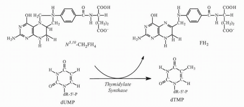
At least two primary mechanisms of action appear capable of causing cell injury: inhibition of TS and incorporation into RNA. FdUMP binds tightly to TS and prevents formation of thymidylate (thymidine 5′-monophosphate, dTMP), the essential precursor of thymidine 5′-triphosphate (dTTP), which is required for DNA synthesis and repair. The functional TS enzyme comprises a dimer of two identical subunits, each of MW ˜30 kDa (bacterial) or ˜36 kDa (human). Each subunit has a nucleotide-binding site and two distinct folate-binding sites, one for 5,10-methylenetetrahydrofolate (5,10-CH2 FH4) monoglutamate or polyglutamate, and one for dihydrofolate polyglutamates. FdUMP competes with the natural substrate 2′-deoxyuridine monophosphate (dUMP) for the TS catalytic site.20,
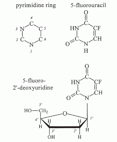
21 During methylation of dUMP, transfer of the folate methyl group to dUMP occurs by elimination of hydrogen attached to the pyrimidine carbon-5 position (
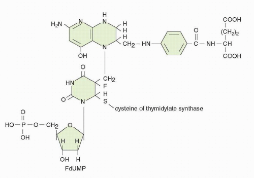
Fig. 9-3). This elimination cannot occur with the more tightly bound fluorine atom of FdUMP, and the enzyme is trapped in a slowly reversible ternary complex with FdUMP and folate (Fig. 9-4). The “thymineless state” that ensues is toxic to actively dividing cells. Toxicity can be circumvented by salvage of dThd in cells that contain dThd kinase. The circulating concentrations of dThd in humans are not thought to be sufficient (approximately 0.1 μmol/L) to afford protection.22 The plasma levels of dThd are approximately tenfold higher in rodents, which complicates preclinical evaluation of the antitumor activity of various TS inhibitors.

A reduced-folate cofactor is required for tight binding of the inhibitor to TS. The natural cofactor for the TS reaction, 5,10-CH2 FH4, in its monoglutamate and polyglutamate forms, binds through its methylene group to the carbon-5 position of FdUMP. The polyglutamates of 5,10-CH2 FH4 are much more effective in stabilizing the FdUMP-TS-folate ternary complex.23 Other naturally occurring folates promote FdUMP binding to the enzyme but form a more readily dissociable complex. Polyglutamated forms of dihydrofolic acid (FH2) promote extremely tight binding of FdUMP to the enzyme.24 FH2 accumulates in cells exposed to methotrexate (MTX). Although MTX is a relatively weak inhibitor of TS in cell-free experiments, MTX polyglutamates are more potent inhibitors.24 MTX polyglutamates decrease the rate of ternary complex formation among FdUMP, folate cofactor, and TS. The ability of MTX polyglutamates to inhibit ternary-complex formation is influenced by the glutamation state of the reduced-folate cofactor and is substantially reduced in the presence of 5,10-CH2 FH4
pentaglutamate.24 Similarly, in tissue culture, MTX-induced depletion of intracellular reduced folates causes a marked reduction in the rate of formation of ternary complex.25


FIGURE 9-3 Thymidylate synthase reaction, with cofactor 5,10 methylene tetrahydrofolate, converting deoxyuridylate (dUMP) to deoxythymidylate (dTMP).

FdUMP binds avidly to the mammalian enzyme, with a dissociation half-life (t1/2) of 6.2 hours.26 Elucidation of the crystal structure of TS has permitted a complex kinetic and thermodynamic description of ternary complex formation.27,28 The interaction proceeds by an ordered mechanism with initial nucleotide binding followed by 5,10-CH2 FH4 binding to form a rapidly reversible noncovalent ternary complex (Fig. 9-4). Enzyme-catalyzed conversions result in the formation of a covalent bond between carbon-5 of FdUMP and the one-carbon unit of the cofactor. The overall dissociation constant of 5,10-CH2 FH4 from the covalent complex is approximately 1 × 10⁻¹¹ mol/L.

Despite the high specificity and potency of TS inhibition by FdUMP and the well-established lethality of dTMP and dTTP depletion, inhibition of TS may not be the sole cause of 5-FU toxicity. If 5-FU toxicity results from dTTP depletion, then dThd should reverse the toxic effects. Examples of complete protection from 5-FU cytotoxicity by dThd have been reported, but dThd shows variable effectiveness in rescuing cells exposed to 5-FU.29 Experimental evidence from in vitro and in vivo studies supports the concept that, depending on the target cell, drug concentration, and modulating factors, 5-FU toxicity may be partially independent of its effect on TS. Coadministration of 5-FU and dThd prevents the early inhibition of DNA synthesis but markedly increases 5-FU toxicity to healthy tissues in the whole animal, increases the antitumor effect of 5-FU against various animal tumors, and increases [3H]FUrd incorporation into RNA.30 Pharmacologic measures that increase FUTP formation and its RNA incorporation also increase its toxicity.


FIGURE 9-4 Interaction of fluorodeoxyuridylate (FdUMP) with thymidylate synthase and N5, 6, 7, 8, 9, 10 CH2 FH4 to form an essentially irreversible complex.


RNA-Directed Effects

5-FU is extensively incorporated into nuclear and cytoplasmic RNA fractions, which may result in alterations in RNA processing
and function, such as inhibiting the processing of initial pre-rRNA transcripts to the cytoplasmic rRNA species in a dose- and time-dependent manner.31, 32, 33, 34

Net RNA synthesis may be inhibited during and after fluoropyrimidine exposure in a concentration- and time-dependent fashion. In some cancer cell lines, a highly significant relationship exists between 5-FU incorporation into total cellular RNA and the loss of clonogenic survival.35 5-FU is incorporated into all species of RNA; substantial amounts of [3H] 5-FU accumulate in low-MW (4S) RNA at lethal drug concentrations.33 Although the analog replaces only a small percentage of uracil residues in RNA, the incorporated 5-FU residues appear to be stable and to persist in RNA for many days after drug administration.36

5-FU exposure affects mRNA processing and translation. Polyadenylation of mRNA and methylation of tRNA are inhibited at relatively low concentrations of 5-FU,37,38 and altered metabolism of specific proteins such as dihydrofolate reductase (DHFR) precursor mRNA has been reported.39 Incorporation of 5-FU into RNA may affect quantitative and qualitative aspects of protein synthesis.40,41

In vitro-transcribed TS mRNA with 100% substitution of 5-FU has an altered secondary structure but no differences in the translational efficiency.40 Hundred percent substitution of uracil residues in human-TS complementary DNA (cDNA) with either FUTP or 5-bromouridine 5′-triphosphate (BrUTP) only inhibited the translational rate in the presence of BrUTP-substituted cDNA.41 The stability of the transcribed mRNA in a cell-free system is increased by threefold and tenfold with FUTP and BrUTP, respectively.

Changes in the structure, levels, and function of small nuclear RNAs (snRNA) and small nuclear ribonuclear proteins (snRNP) result from 5-FU treatment.42,65 The substitution of FUTP for uridine triphosphate (UTP) in a cell-free system (84% replacement of uracil residues by 5-FU) leads to pH-dependent missplicing of [32P]-labeled human β-globin precursor mRNA; pH values favoring 5-FU ionization promote missplicing.43

Further, 5-FU substitution greatly increases the pH and temperature sensitivity of the process. Partial ionization of 5-FU residues at physiologic pH (pK 5-FU = 7.8 versus pK uracil = 10.1) may therefore destabilize the active conformation of RNA.44

Another potential locus of 5-FU action is inhibition of enzymes involved in posttranscriptional modification of RNA particularly the formation of methylated uracil bases that have profound effects on splicing.45a,45b, 46, 47 Although 5-FU-associated cytotoxicity in cancer cells exposed in the presence of sufficient concentrations of dThd to circumvent TS inhibition is presumed to result from RNA-directed effects of 5-FU, it is paradoxical that significant incorporation of 5-FU into RNA may occur in some cancer cell lines in the absence of toxicity. The factors that influence whether 5-FU-RNA incorporation results in cytotoxicity are not clear. The rate of RNA incorporation and the species into which the fluoropyrimidine is incorporated may be more important determinants of cytotoxicity than the total amount incorporated. 5-FU and FUrd may be channeled into different ribonucleotide compartments and, ultimately, into distinct classes of RNA.48

In summary, the changes that result in altered pre-RNA processing and mRNA metabolism are not uniform for all RNA species after 5-FU exposure. Effects on precursor and mature rRNA, precursor and mature mRNA, tRNA, and snRNA species suggest inhibition of processing; incorporated 5-FU residues also inhibit enzymes involved in posttranscriptional modification of uracil. Many of the RNA-directed effects of 5-FU undoubtedly occur as a consequence of its fraudulent incorporation into various RNA species. The changes in certain key mRNAs resulting from 5-FU exposure may be relevant to its cytotoxicity. 5-FU-mediated interference with the production of enzymes involved in DNA repair may have cytotoxic consequences, such as 5-FU-mediated inhibition of ERCC-1 mRNA expression in cisplatin-resistant cancer cells.49


DNA-Directed Mechanisms of Potential Toxicity

The biochemical consequences of TS inhibition and the potential effects on DNA integrity have been extensively studied, but their relationship to cytotoxicity is incompletely understood. Inhibition of TS results in depletion of dTMP and dTTP, thus leading to inhibition of DNA synthesis and interference with DNA repair. Accumulation of dUMP occurs behind the blockade of TS, and further metabolism to the deoxyuridine triphosphate (dUTP) level may occur.50 Inhibition of TS is accompanied by elevated concentrations of deoxyuridine in the extracellular media in cell culture models and in plasma of rodents; monitoring changes in plasma deoxyuridine levels may, therefore, serve as an indirect reflection of TS inhibition.

FdUTP and dUTP are substrates for DNA polymerase, and their incorporation into DNA is a possible mechanism of cytotoxicity.51, 52, 53, 54 5-FU cytotoxicity in some models correlates with the level of 5-FU-DNA.52,53 Two mechanisms prevent incorporation of FdUTP and dUTP into DNA. The enzyme dUTP pyrophosphatase or dUTP hydrolase catalyses the hydrolysis of FdUTP to FdUMP and inorganic pyrophosphate.55,56 The DNA repair enzyme uracil-DNA-glycosylase hydrolyzes the 5-FU-deoxyribose glycosyl bond of the FdUMP residues in DNA, thereby creating an apyrimidinic site.57 The base deoxyribose 5′-monophosphate is subsequently removed from the DNA backbone by an AP (apurinic/apyrimidinic) endonuclease, creating a single-strand break, which is subsequently repaired. With dThd triphosphate depletion, however, the efficiency of the repair process is substantially weakened. Uracil-DNA-glycosylase is a cell cycle-dependent enzyme with maximal levels of activity at the G1 and S interface, such that excision of the fraudulent bases occurs before DNA replication. The activity of uracil-DNA-glycosylase inversely correlates with the level of FdUrd incorporation into DNA in human lymphoblastic cells. Because the affinity of human uracil-DNA-glycosylase is much lower for 5-FU than for uracil, 5-FU is removed more slowly from DNA by this mechanism.57 FdUTP itself inhibits the activity of uracil-DNA-glycosylase.58 Accumulation of deoxyadenosine triphosphate (dATP) accompanies TS inhibition.59 The combined effects of deoxyribonucleotide imbalance (high dATP, low dTTP, high dUTP) and misincorporation of FdUTP into DNA may have deleterious consequences affecting DNA synthesis, the integrity of nascent DNA, and induction of apoptosis.

A variety of DNA-directed effects have been described.60, 61, 62, 63, 64 5-FU treatment inhibits DNA elongation and decreases the average DNA chain length.60 DNA strand breaks accumulate in 5-FU-treated cells and correlate with excision of [3H]5-FU from DNA. 5-FU and FdUrd result in single- and double-stranded DNA breaks in HCT-8 cells in a concentration- and time-dependent fashion, a process that is enhanced by LV and limited by dThd.61 FdUrd
exposure may result in the formation of large (1 to 5 Mb) DNA fragments as a result of double-strand DNA breaks; the time course and extent of DNA megabase fragmentation correlate with loss of clonogenicity in HT29 cells.62 The pattern of DNA fragmentation is distinct from that associated with γ radiation, which produces random breaks. The pattern of high-MW DNA damage differs in SW620 cells, which are equally sensitive as HT29 cells to FdUrdinduced inhibition of TS, but require higher drug concentrations and longer exposures to achieve a comparable degree of DNA fragmentation and cytotoxicity. Resistance to 5-FU appears to be related to the higher activity of dUTPase and failure to accumulate dUTP of SW620 cells.63 Simple dThd starvation of a TS-deficient murine cell line produces much smaller DNA fragments, 50 to 200 kb in length.64

Inhibition of protein synthesis by cycloheximide within 8 hours of FdUrd exposure dramatically reduces DNA double-strand breakage and lethality in murine FM3A cells, suggesting that FdUrd exposure triggers the synthesis of an endonuclease capable of inducing DNA strand breaks.60 Factors that regulate recognition of DNA damage and apoptosis contribute to 5-FU lethality. The oncogene p53 plays a pivotal role in the regulation of cell-cycle progression and apoptosis and influences the sensitivity of murine embryonic fibroblasts to 5-FU.65 Transfection and expression of the bcl-2 oncogene in a human-lymphoma cell line render it resistant to FdUrd. TS inhibition, dTTP depletion, and induction of single-strand breaks in nascent DNA are similar in vector control cells and bcl-2-expressing cells.66 In vector control cells, induction of double-stranded DNA fragmentation in parental DNA coincides with onset of apoptosis. The contribution of DNA damage to cell lethality varies among different malignant lines, and DNA fragmentation does not appear to contribute to 5-FU-mediated cytotoxicity in some cancer cell lines.67

In summary, TS inhibition, as seen in “pure” form with FdUrd treatment in the absence of dThd salvage, and 5-FU incorporation into RNA are capable of producing lethal effects on cells. DNA damage also contributes to cytotoxicity and can occur in the absence of detectable FdUTP incorporation into DNA. The combined effects of deoxyribonucleotide imbalance (high dATP, low dTTP, high dUTP) and misincorporation of FdUTP and dUTP into DNA result in deleterious consequences affecting DNA synthesis and the integrity of nascent DNA. The pattern and extent of DNA damage induced by fluoropyrimidines in human colorectal cancer cells vary and may be affected by the activity of enzymes involved in DNA repair and by downstream pathways that are required to implement cellular destruction.

It is now recognized that the genotoxic stress resulting from TS inhibition activates programmed cell-death pathways, resulting in induction of parental DNA fragmentation. Depending on the cell line in question, two different patterns of parental DNA damage may be noted: internucleosomal DNA laddering, the hallmark of classical apoptosis, and high-MW DNA fragmentation with segments ranging from approximately 50 kb to 1 to 3 Mb. Differences in the type and activity of endonucleases and DNA-degradative enzymes triggered in a given cell line most likely explain these disparate patterns of parental DNA fragmentation. In “apoptosis-competent” cancer cell lines, such as HL60 promyelocytic leukemia cells, genotoxic stress results in rapid (within hours) induction of programmed cell death, with classic DNA laddering. In contrast, many cancer cell lines derived from epithelial tumors, including colon cancer, appear to undergo delayed programmed cell death. This phenomenon may reflect a “postmitotic” cell death, in which one or more rounds of mitosis are needed before cell death occurs.68 In such cell lines, the duration of the genotoxic insult may determine whether induction of cytostasis or programmed cell death occurs. One possible explanation for delayed apoptosis is that originally sublethal damage to genes, essential for cell survival, may ultimately lead to cell death with subsequent rounds of DNA replication.

Factors operating downstream from TS clearly influence the cellular response to genotoxic stress, such as overexpression of the cellular oncoproteins bcl-2 and mutant p53. Disruption of the signal pathways that sense genotoxic stress or lead to induction of programmed cell death, or both, may render a cancer cell inherently resistant to 5-FU. In some cancer cell lines, thymineless death may be mediated by Fas and Fas-ligand interactions.69 Cancer cell lines insensitive to Fas-mediated apoptosis are insensitive to 5-FU, suggesting that modulation of their expression may influence sensitivity to 5-FU.70 As previously mentioned, base excision repair (BER) plays an essential role in removing incorporated 5-FU and uracil residues from DNA, resulting in single-strand DNA breaks. BER recognizes the mispairing of 5-FU with guanine in DNA and excises the fluoropyrimidine. Because BER involves multiple proteins, deficiencies in one of the components, such as uracil DNA glycosylase, XRCC1 or DNA polymerase-β, may reduce the toxic effects of TS inhibitors.71 Abrogation of uracil DNA glycosylase activity affords protection from cytotoxicity in selected cell lines72,73 but not others.73

Microsatellite instability (MSI) is a manifestation of genomic instability in human cancers that have a decreased overall ability to faithfully replicate DNA and is a surrogate phenotypic marker of underlying functional inactivation of the human DNA mismatch repair (MMR) genes.74 In vitro studies suggest that MMR recognizes DNA breaks resulting from 5-FU incorporated into DNA and signals cell cycle (G2M) arrest. Functional loss of a MMR gene results from inactivation of both alleles via some combination of coding region mutations, loss of heterozygosity, and/or promoter methylation, which leads to gene silencing. In vitro studies suggest that MMR-proficient cells are more sensitive to 5-FU or FdUrd than MMR-deficient cells.75,76 The loss of MMR proficiency leads to fluoropyrimidine resistance in some cell lines, but not others,77 perhaps because of differences in cell lines and conditions of drug concentration and duration of exposure in vitro. The MSI phenotype has been associated with a better prognosis in stage-for-stage matched tumors in primary colorectal cancer,78 but data are conflicting as to whether MSI status influences benefit from 5-FU-based adjuvant therapy. The MSI phenotype has been associated with a better prognosis in stage for stage-matched tumors in primary colorectal cancer. While there is conflicting evidence for the predictive effect of the MSI phenotype in stage 3 and 4 patients, there is an emerging consensus that stage 2 patients with an MSI high phenotype do not gain any benefit from adjuvant 5-FU therapy and may actually be harmed by 5-FU treatment through undetermined mechanisms.392 An ongoing prospective trial in stage 2 colon cancer patients will help clarify this issue.


Relative Importance of RNA- Versus DNA-Directed Effects

The relative contributions of DNA- and RNA-directed mechanisms to the cytotoxicity of 5-FU are influenced by the specific patterns of intracellular drug metabolism, which vary among different healthy and tumor tissues. 5-FU concentration and duration of exposure play pivotal roles in determining the basis of cytotoxicity. The improved response rates observed with LV modulation of bolus 5-FU therapy, the correlation between high TS expression in tumor tissue and insensitivity to 5-FU-based therapy, and the clinical activity of the antifolate-based TS inhibitors provide strong evidence that TS is the most important therapeutic target. In some models, RNA-directed effects have been predominant, particularly with prolonged duration of exposure, and are not necessarily cell-cycle dependent, whereas DNA-directed effects have been important during short-term exposure of cells in S phase. However, this generalization does not hold for all experiments with selected tumor cell lines.79

In two human colon carcinoma cell lines, the determinants of cytotoxicity with prolonged (120-hour) exposure to 5-FU at pharmacologically relevant concentrations (0.1 to 1.0 μmol/L) suggested that DNA-directed effects (inhibition of TS and induction of single-strand breaks in nascent DNA) and the gradual and stable accumulation of 5-FU into RNA both contribute to 5-FU toxicity.80 Thus, the primary mechanism of 5-FU cytotoxicity varies among cancer cell lines and can change within a given cell line by alterations in schedule or the circumstances of drug exposure (the presence or absence of potential modulators of toxicity). More than one mechanism of action may be operative, and each may contribute to cytotoxicity.


Determinants of Sensitivity to Fluoropyrimidines

Because of the complexity of fluoropyrimidine metabolism and the multiple sites of biochemical action, multiple factors may be associated with responsiveness to this class of antimetabolites (Table 9-2). Deletion of or diminished activity of the various activating enzymes may result in resistance to 5-FU.81, 82, 83 Conversely, elevated levels of certain activating enzymes have been associated with increased fluoropyrimidine sensitivity. Clones derived from murine leukemia selected for stable resistance to either 5-FU, FUrd, or FdUrd are each deficient in one enzyme involved in pyrimidine metabolism: decreased OPRT was associated with 5-FU resistance, whereas FdUrd and FUrd resistance were associated with deletion of dThd and Urd kinase, respectively.84,85 Clones retained sensitivity to alternate fluoropyrimidines; thus, resistance to 5-FU may not preclude sensitivity to FdUrd, or vice versa.

In addition to the importance of these activating enzymes, the availability of ribose-1-phosphate, dR-1-P, and PRPP may influence activation and response.84,85 Guanine nucleotides augment 5-FU activation to the ribonucleotide and deoxyribonucleotide levels by serving as a source of ribose-1-phosphate and dR-1-P.

The formation of 5-fluoropyrimidine nucleotides within target cells and the size of the competitive physiologic pools of UTP and dTTP also influence 5-FU cytotoxicity.86,87 The extent of 5-FU incorporation into RNA depends on FUTP formation and the size of the competing pool of UTP. Strategies that increase FUTP formation generally increase incorporation of FUTP into RNA and enhance 5-FU toxicity. Modulators including 6-methylmercaptopurine riboside (MMPR), N-phosphonoacetyl-L-aspartic acid (PALA), pyrazofurin, MTX, and dThd may increase FUTP formation by virtue of inhibiting de novo purine or pyrimidine synthesis, thereby increasing PPRP levels. Through feedback inhibition, expansion of dTTP pools decreases FdUMP formation by two means: blocking phosphorylation of FdUrd by dThd kinase and inhibiting the reduction of FUDP to FdUDP. In contrast, expansion of UTP or cytidine triphosphate (CTP) pools inhibits formation of fluorouridine monophosphate (FUMP) by Urd kinase. Changes in nucleotide pool size have been implicated in 5-FU resistant murine S49 lymphoma sublines that increased CTP synthase activity, increased CTP pools, and decreased UTP pools.88


TABLE 9.2 Determinants of sensitivity to 5-FU


Extent of 5-FU anabolism to FdUMP and triphosphate nucleotides


Activity of anabolic enzymes


Availability of (deoxy)ribose-1-phosphate donors and phosphoribosyl phosphate


Activity of catabolic pathways


Alkaline and acid phosphatases


Dihydropyrimidine dehydrogenase


Thymidylate synthase


Baseline activity of TS


Intracellular reduced-folate content


Polyglutamation of folate cofactor


Concentration of competitive dUMP


Upregulation of TS protein expression or TS amplification or mutation


Extent of fluorouridine triphosphate incorporation into RNA


Concentration of competing normal substrates (UTP)


Extent of dUTP and FdUTP incorporation into DNA


Ability to increase pools of dUTP (dUTP hydrolase activity)


Uracil-DNA-glycosylase activity


Extent and type of DNA damage


Single-strand breaks


Double-strand breaks


Newly synthesized DNA versus parental DNA


DNA repair


Cellular response to genotoxic stress


Cytostasis and repair of DNA versus cell death


Intact programmed cell death signaling pathways


Duration of genotoxic stress


FdUrd, 5-fluoro-2′-deoxyuridine; FUrd, 5-fluoro-uridine; dUMP, deoxyuridine monophosphate; FdUMP, 5-fluoro-2′deorguridine monophosphate; TS, thymidylate synthase; 5-FU, 5-flurouracil; dUTP, deoxyuridine triphosphate; FdUTP, dUTP deoxyuridine triphosphate; UTP, uridine triphosphate.


Because RNA- and DNA-directed effects of 5-FU may differ in importance among different malignant cell lines, any single manipulation of 5-FU metabolism may produce conflicting results if different tumor models are compared. The development and
application of sensitive assays that permit reliable measurement of FUTP, 5-FU-RNA levels, and TS inhibition in patient samples will help elucidate clinical determinants of sensitivity to fluorinated pyrimidines given by various schedules. In one study, RNA and DNA incorporation in tumor biopsy specimens taken 2, 24, or 48 hours from patients receiving bolus 5-FU (500 mg/m2) were measured using gas chromatography/mass spectrometry (GC/MS) after complete degradation of isolated RNA and DNA to bases. Maximal incorporation occurred 24 hours after 5-FU administration: 1.0 pmol/mg RNA (n = 59) and 127 pmol/mg DNA (n = 46). Incorporation into RNA, but not DNA, significantly correlated with intratumoral 5-FU levels. The extent of TS inhibition, but not RNA or DNA incorporation, correlated with response to 5-FU therapy.89 Results of such studies in clinical samples from patients receiving various infusional schedules of 5-FU are needed for a fuller understanding of determinants of response.


Determinants of Thymidylate Synthase Inhibition

The ability of FdUMP to inhibit TS is influenced by several variables, including the concentration of enzyme, the amount of FdUMP formed and its rate of breakdown, the levels of the competing healthy substrate (dUMP) and 5,10-CH2 FH4 cofactor, and the latter’s extent of polyglutamation. The degree and persistence of TS inhibition are a crucial determinant of cytotoxicity. Blockade of TS can lead to a gradual expansion of the intracellular dUMP pool; resumption of DNA synthesis is a function of three factors: the rate of decrease of intracellular FdUMP, the rate of increase in dUMP, which competes with FdUMP for newly synthesized TS and for enzyme that has dissociated from the ternary complex, and the rate of synthesis of new TS.

FdUMP accumulates rapidly in both responsive L1210 leukemia and resistant Walker 256 carcinoma, but more rapid recovery of DNA synthesis in the insensitive line correlates with accelerated decline in intracellular free FdUMP concentrations.86 The basis for resistance in some cells may be explained by the rate of nucleotide inactivation rather than slower formation of the active product.

Determination of TS content in tumor tissue may help to clarify the relationship between pretreatment TS levels and prognosis, response, or both, to 5-FU therapy. Biochemical assays permit measurement of dUMP, TS, the ternary complex, and free FdUMP in tissue samples.87,90,91 Their application to clinical tumor samples is limited by the need for relatively large quantities of tissue (at least 50 mg) as well as fresh or frozen tumor tissue. In one study, biopsies of liver metastases were obtained 20 to 240 minutes after 500 mg/m2 5-FU among 21 patients undergoing elective surgery, and maximal TS inhibition occurred within 90 minutes and averaged 70% to 80% in tumor tissue.92 Large variations in TS binding and catalytic activity were noted in primary colon tumors, but the overall enzyme levels were significantly higher than in adjacent healthy colonic tissue.93

Measurement of TS gene expression provides an alternative to directly assaying intracellular TS enzyme. Overexpression to TS in tumor biopsies correlates with insensitivity to 5-FU-based regimens.94,95

Monoclonal antibodies have been developed that are capable of detecting human TS in immunoprecipitation and enzyme-linked immunosorbent assays (ELISA) and by immunoblot analysis. A number of studies have reported a strong relationship between TS expression in clinical specimens and prognosis; a systematic review of such studies in colorectal cancer has found great variability in methodology, clinical regimens, and combinations of drugs employed and concluded that high TS expression had the most consistent correlation with decreased survival in patients with metastatic disease,96 although defective mismatch repair was also implicated in an improved over-all prognosis but a poorer response to 5-FU.97

Quantitative and qualitative changes in TS have been identified in cells with innate or acquired resistance to fluoropyrimidines. Amplification of the TS gene, with corresponding elevation of enzyme content, has been found in lines resistant to 5-FU or FdUrd.98, 99, 100 Resistant cell lines may have an altered TS protein with either decreased binding affinity for FdUMP or decreased affinity for 5,10-CH2 FH4.101, 102, 103, 104 Adequate reduced-folate pools are required to form and maintain a stable ternary complex. Administration of exogenous reduced folates enhances the cytotoxicity of 5-FU and FdUrd in preclinical models, and clinical administration of LV is used to elevate the reduced-folate content in the cancer cell.105,106 Tumor cells transport LV intracellularly and convert the folates to more potent and stable polyglutamates. Deficiency of the low-affinity, high-capacity folate transport system (impaired membrane transport) or reduced folylpolyglutamate synthetase activity (impaired polyglutamation) would likely impair the ability of LV to expand the reduced-folate pools, although their mechanisms of resistance have not been found in experiments to date.

In summary, to inhibit TS, 5-FU must enter the tumor and then be metabolized to FdUMP. Additional factors influence the ability of FdUMP to inhibit TS, including enzyme concentration, mutations affecting TS binding, and cofactor concentration. The tumor cell must enter the vulnerable synthetic phase of the cell cycle during drug exposure. A final factor, the ratio of endogenous dUMP to FdUMP pools can affect the duration of TS inhibition.


Regulation of Thymidylate Synthase

TS is required for DNA replication; its activity is higher in rapidly proliferating cells than in noncycling cells. When nonproliferating cells are synchronized and stimulated to enter the synthetic phase of the cell cycle, TS content may increase up to 20-fold.107 In proliferating cancer cells, TS activity varies by fourfold to eightfold from resting to synthetic phase.108 Increased expression of the TS gene at the G1-S boundary is controlled by both transcriptional and posttranscriptional regulation.109,110 Elements in the promoter region of the human TS gene are regulated by the transcription factor LSF, which is in turn controlled by the astrocyte elevated gene-1 (AEG-1) AEG-1, when overexpressed in hepatocyte Uular carcinoma cells, up-regulates TS and dihydropyrimidine dehydrogenase (DPD), and confers resistance to 5-FU.110

In both experimental and clinical111,112 observations,113 5-FU exposure may be accompanied by an acute increase in TS content, which may in turn permit recovery of enzymatic activity, and the magnitude of the increase is influenced by drug concentration and time of exposure. In NCI-H630 colon cancer cells, TS content increases up to 5.5-fold during 5-FU exposure and is regulated at the translational level.114


TS protein binds to specific regions in its corresponding TS-mRNA, which contributes to the regulation of TS-mRNA translation.115,116 Antisense oligodeoxynucleotides targeted at the AUG translational start site of TS-mRNA inhibit translation in rabbit reticulocyte lysate; transfection of KB31 nasopharyngeal cancer cells with a plasmid construct containing the TS antisense fragment decreases the expression of TS protein and enhances the sensitivity to FdUrd by eightfold.117 Reduced-folate content also influences TS expression. TS (TS-C1) with reduced affinity for its folate cofactor has normal clonogenic growth in the presence of high folate levels, suggesting folate responsiveness of the TS-C1 mutant.118 Exposure of TS-C1 cells to 20 μmol/L LV stimulated de novo dTMP synthesis, whereas TS activity was lost by 24 hours after LV removal.118


Importance of Schedule of Administration in Preclinical Models

Drug concentration and duration of exposure in vitro are important determinants of response to 5-FU.29,36,119, 120, 121 High drug concentrations (above 100 μmol/L) are generally required for cytotoxicity if the duration of exposure is brief (<6 hours), whereas prolonged exposure (>72 hours) to concentrations between 1 and 10 μmol/L effectively kills many tumors in culture. Schedules designed to provide extended exposure are currently preferred in current clinical regimens, usually in combination with LV.

Other molecular pathways undoubtedly modulate fluoropyrimidine toxicity. Loss of protein expression of pRB (the retinoblastoma protein which controls cell cycle entry and proliferation rates) correlates with sensitivity to FU and to MTX in mouse embryo fibroblasts and in selected breast and colon cancer cell lines,122 and the same correlation was found for pRB expression in tumor samples from breast cancer patients treated with adjuvant cyclophosphamide, 5-FU, and MTX. However, the number of patients with tumors lacking pRB immunostaining was very small (<5%). The authors hypothesize that the lack of cell cycle control allowed cells to proceed through DNA synthesis while accumulating DNA damage due to drug treatment.

FU treatment activates the MAP kinase pathway and the downstream Egr1 transcription factor, and thereby stimulates production of thrombospondin-1, an antiangiogenic cytokine. This finding leads to speculation that fluoropyrimidines may synergize with inhibitors of the vascular endothelial growth factor (VEGF) pathway,123 as has been observed clinically with bevicizumab.


Clinical Pharmacology of 5-Fluorouracil

The pharmacokinetics of 5-FU are important because of the choices of routes and schedules of administration available for this drug. Regional approaches permit selective exposure of specific tumor-bearing sites to high local concentrations of drug. Pharmacokinetic studies have played an important role in assessing these alternative schedules and routes of administration.


Clinical Pharmacology Assay Methods

5-FU has been assayed in biologic fluids using high-performance liquid chromatography (HPLC) and GC-MS. In general, an initial deproteination step is performed by chemical or filtration techniques. HPLC methods using ultraviolet detection of 5-FU are typically associated with limits of detection in the range of 0.2 to 1.0 μmol/L. Column or valve-switching techniques and the use of microbore-HPLC columns can further improve the limits of detection. Radio immuno assays of 5-FU, with high specificity and sensitivity, have also been developed.

The nucleoside metabolites of 5-FU can be separated from parent drug on reversed-phase and ion-exchange columns, whereas separation of the nucleotide metabolites is obtained with either anion-exchange or reversed-phase ion-pairing methods. Preclinical studies describing intracellular metabolism generally typically use radiolabeled 5-FU; HPLC with inline liquid scintillation detection is used to quantify the metabolites.

Derivatization of 5-FU is required for GC-MS. MS generally provides much greater sensitivity than that achievable with HPLC, with limits of detection as low as 0.5 ng/mL (4 nmol/L) for a 1-mL plasma sample.124,125 Recent advances in fluorine-19 magnetic resonance imaging (MRI) have permitted monitoring of the pharmacokinetics and cellular pharmacology of 5-FU, thus allowing noninvasive determination of 5-FU content in tissues.126

5-FU is unstable in whole blood and plasma at room temperature, and catabolism is much more rapid in whole blood than in plasma.127 Blood samples should be placed on ice immediately; plasma should be quickly isolated. 5-FU is stable in plasma at 4°C for up to 24 hours and is stable for prolonged periods when stored at −20°C.


Absorption and Distribution

Bioavailability of 5-FU by the oral route is highly variable. Less than 75% of a dose reaches the systemic circulation.128 When administered by intravenous bolus or infusion, 5-FU readily penetrates the extracellular space, cerebrospinal fluid (CSF), and extracellular “third-spaces” such as effusions. The volume of distribution (Vd) ranges from 13 to 18 L (8 to 11 L/m2) after intravenous bolus doses of 370 to 720 mg/m2, which slightly exceeds extracellular fluid space.124,129


Plasma Pharmacokinetics

The pharmacokinetic profile of 5-FU varies according to dose and schedule of administration. After intravenous bolus injection of 370 to 720 mg/m2, peak plasma concentrations (Cp) of 5-FU vary widely and primarily lie in the range of 300 to 1,000 μmol/L (Table 9-3).124,129, 130, 131 Rapid metabolic elimination accounts for a primary t1/2 of 8 to 14 minutes; 5-FU Cp fall below 1 μmol/L within 2 hours.

The most sensitive assays detect triexponential elimination of intravenous bolus 5-FU with t1/2 values of 2, 12, and 124 minutes.132 A prolonged third elimination phase of 5-FU was noted by GC-MS after bolus administration with a t1/2 of 5 hours: 5-FU plasma concentration ranged from 36 to 136 nmol/L 4 to 8 hours after intravenous bolus doses of 500 to 720 mg/m2 and may reflect tissue release.124

The clearance of 5-FU is much faster with continuous infusion (CI) (Table 9-4) than with bolus administration and increases as the dose rate decreases (Fig. 9-5).125,128,133, 134, 135, 136, 137, 138 As the duration of 5-FU infusion increases, the tolerated daily dose decreases. A recommended starting dose of single-agent 5-FU given by protracted CI is 300 mg/m2; the achieved steady-state plasma levels (Css) are in the submicromolar range. With CI over 96 to 120 hours, a daily dose of 1,000 mg/m2 produces a Css in the 1 to 3 μmol/L range, and an intermittent schedule is necessary. CI of 2,000 to 2,600 mg/m2 5-FU daily given either for 72 hours every 3 weeks or for 24 hours weekly yields a Css of 5 to 10 μmol/L.


TABLE 9.3 Pharmacokinetics of 5-FU given by intravenous bolus


Investigators


Dose (mg/m2)


No.


Half-life (min)


Clearance (mL/min/m2)


Plasma concentration (μmol/L)


AUC per dose (μmol/min/L)


Grem et al.129


370


16


8.1 ± 0.4


862 ± 24


C0: 332 ± 27


3,761 ± 286


15 min: 82 ± 6


60 min: 4 ± 1


Macmillan et al.152


400


8


11.4 ± 1.5


744 ± 145


5 min: 469 ± 85


9,885 ± 1,569


20 min: 100 ± 20


60 min: 13 ± 6


Heggie et al.130


500


10


12.9 ± 7.3


594 ± 7.3


5 min: 420 ± 102


7,125 ± 2,371


20 min: 114 ± 52


60 min: 10 ± 11


van Gröeningen et al.124


500


15


9.8 ± 2.4


558


Not stated


7,338 ± 1,708


600


18


404


12,000 ± 2,446


720


7


14.4


± 2.5


349


16,200 ± 2,446


Grem et al.131


425


11


9.8 ± 0.5 (all doses)


743 ± 81


C0: 378 ± 46


4,401 ± 363


490


13


713 ± 28


393 ± 24


5,304 ± 227


AUC, area under the concentration time curve; C0, estimated initial concentration.


Note: If either AUC or clearance was not provided, it was calculated from the following equation: intravenous dose/AUC = clearance. The MW of 5-FU = 130.1.


TABLE 9.4 Pharmacokinetics of 5-FU given by continuous intravenous infusion


Investigators


Duration of infusion


Daily dose (mg/m2)


No.


Css (μmol/L)


Clearance (mL/min/m2)


Grem et al.136


Protracted


64-200


24


0.30 ± 0.04 (0.14-1.04)


3,050 ± 330


Anderson et al.125


Protracted


176-300


3


0.32 (0.05-0.57)


Not provided


Harris et al.135


Protracted


300


7


0.13 ± 0.01


Not provided


Yoshida et al.134


Protracted


190-600


19


1.15 ± 0.15 (0.08-2.40)


2,033


Petit et al.137


120 h


450-966


7


2.6 ± 0.2


Not provided


Fleming et al.138


120 h


1,000


57


2.1


2,523 ± 684


Fraile et al.128


96 h


1,000-1,100


6


24-48 h, 1.3 ± 0.1


Not provided


72-96 h, 1.8 ± 0.3


Benz et al.133


24 h


1,500


7


4 (1.94-5.63)


2,118 (1,235-3,471)


Erlichman et al.139


120 h


1,250


15


3.4 ± 0.4


2,410


1,500


6


5.1 ± 1.0


1,790


1,750


14


6.4 ± 0.9


1,990


2,000


25


7.2 ± 0.7


1,910


2,250


17


7.5 ± 1.0


2,000


Remick et al.140


72 h


1,655


6


5.4 ± 0.3


1,750 ± 105


2,875


8


13.9 ± 0.5


1,117 ± 37


Grem et al.141


72 h


1,150-1,525


19


3.4 ± 0.5


3,011 ± 356


1,750


31


5.0 ± 0.5


2,671 ± 563


2,000


53


6.5 ± 0.9


2,651 ± 324


2,300


14


8.8 ± 1.3


2,116 ± 572


2,645


10


10.0 ± 2.1


2,247 ± 443


Css, plasma concentration at steady state.


Note: Plasma clearance converted from milliliter per minute assuming an average body surface area of 1.7 m2 and from milliliter per kilogram assuming a conversion factor of 37 from kg to m2.


FIGURE 9-5 Relationship of 5-FU infusion rate to 5-FU steady-state concentration in plasma. (Adapted from data in Table 7-6, 4th ed. Principles and Practice of Cancer Chemotherapy and Biological Response Modifiers.)

5-FU clearance varies considerably between individuals. The elimination kinetics of 5-FU are nonlinear.124,132, 133, 134, 135, 136, 137, 138, 139, 140, 141, 142, 143, 144 The following are noted with increasing doses: a decrease in hepatic extraction ratio; an increase in oral bioavailability; an increase in plasma t1/2; a decrease in total-body clearance; and an increase in 5-FU area under the curve (AUC). Although the change in 5-FU clearance or AUC with increasing 5-FU dosage on a given schedule may be linear over a certain dose range, with higher dosages these parameters may change disproportionately. This nonlinear behavior represents saturation of metabolic processes at higher drug concentrations, leading to difficulty in predicting plasma levels or toxicity at higher dosages.

Variation in 5-FU pharmacokinetics has been reported to vary up to fivefold according to time of day but two studies found virtually opposite times of day for peak and trough value.135,137 This discrepancy suggests that other factors may have influenced rates of 5-FU clearance.

5-FU pharmacokinetics have correlated with toxicity,124,129,134,136,138, 139, 140, 141, 142, 143, 144, 145, 146, 147 as reflected in the total AUC with bolus injection,131 or with plasma concentration of drug at steady state in patients receiving treatment by CI.147 Dose adjustment based on pharmacokinetics appears to be useful in avoiding clinical toxicity147, 148

Only gold members can continue reading. Log In or Register to continue

Stay updated, free articles. Join our Telegram channel

May 27, 2016 | Posted by in ONCOLOGY | Comments Off on 5-Fluoropyrimidines

Full access? Get Clinical Tree

Get Clinical Tree app for offline access